

(N) and actuator domain (A) [91]. This case study shows the selectivity gain by
bound inhibitor, utilizing the domain flexibility of receptor [94].
2.5 Effect of pH on Binding Affinities
Protonation states of the titratable groups participating in the binding can have
significant effect on the binding affinity of the interaction [16]. Waelbroeck [95]
presented a model with assumptions that correct ionization state of all active groups
is the requisite for binding, and ionization state of non-binding residues does not
affect binding to study quantitative effect of pH change on binding affinity of the
receptor–ligands interaction. They chose pH dependency of insulin and insulin
analogs binding to their cellular receptor to study their model [95].
logðKÞ ¼ log K real
ð
Þþ log R
Ã
=R
ð
Þþ log L
Ã
=L
ð
Þ
ð1Þ
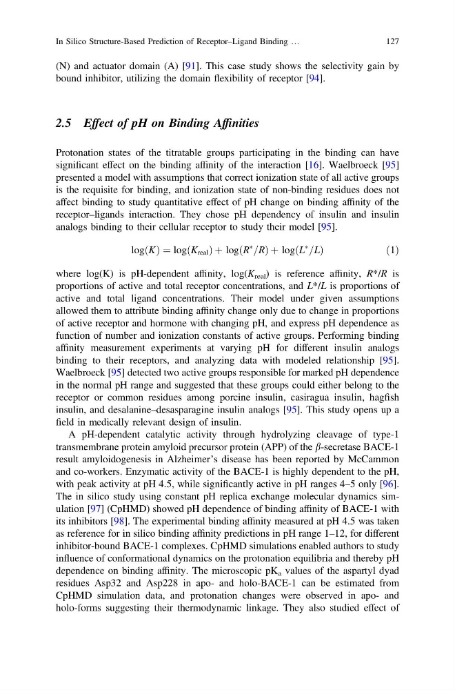
where log(K) is pH-dependent affinity, log(K real ) is reference affinity, R*/R is
proportions of active and total receptor concentrations, and L*/L is proportions of
active and total ligand concentrations. Their model under given assumptions
allowed them to attribute binding affinity change only due to change in proportions
of active receptor and hormone with changing pH, and express pH dependence as
function of number and ionization constants of active groups. Performing binding
affinity measurement experiments at varying pH for different insulin analogs
binding to their receptors, and analyzing data with modeled relationship [95].
Waelbroeck [95] detected two active groups responsible for marked pH dependence
in the normal pH range and suggested that these groups could either belong to the
receptor or common residues among porcine insulin, casiragua insulin, hagfish
insulin, and desalanine–desasparagine insulin analogs [95]. This study opens up a
field in medically relevant design of insulin.
A pH-dependent catalytic activity through hydrolyzing cleavage of type-1
transmembrane protein amyloid precursor protein (APP) of the b-secretase BACE-1
result amyloidogenesis in Alzheimer’s disease has been reported by McCammon
and co-workers. Enzymatic activity of the BACE-1 is highly dependent to the pH,
with peak activity at pH 4.5, while significantly active in pH ranges 4–5 only [96].
The in silico study using constant pH replica exchange molecular dynamics simulation [97] (CpHMD) showed pH dependence of binding affinity of BACE-1 with
its inhibitors [98]. The experimental binding affinity measured at pH 4.5 was taken
as reference for in silico binding affinity predictions in pH range 1–12, for different
inhibitor-bound BACE-1 complexes. CpHMD simulations enabled authors to study
influence of conformational dynamics on the protonation equilibria and thereby pH
dependence on binding affinity. The microscopic pK a values of the aspartyl dyad
residues Asp32 and Asp228 in apo- and holo-BACE-1 can be estimated from
CpHMD simulation data, and protonation changes were observed in apo- and
holo-forms suggesting their thermodynamic linkage. They also studied effect of
In Silico Structure-Based Prediction of Receptor–Ligand Binding …
127
bound inhibitor, utilizing the domain flexibility of receptor [94].
2.5 Effect of pH on Binding Affinities
Protonation states of the titratable groups participating in the binding can have
significant effect on the binding affinity of the interaction [16]. Waelbroeck [95]
presented a model with assumptions that correct ionization state of all active groups
is the requisite for binding, and ionization state of non-binding residues does not
affect binding to study quantitative effect of pH change on binding affinity of the
receptor–ligands interaction. They chose pH dependency of insulin and insulin
analogs binding to their cellular receptor to study their model [95].
logðKÞ ¼ log K real
ð
Þþ log R
Ã
=R
ð
Þþ log L
Ã
=L
ð
Þ
ð1Þ
where log(K) is pH-dependent affinity, log(K real ) is reference affinity, R*/R is
proportions of active and total receptor concentrations, and L*/L is proportions of
active and total ligand concentrations. Their model under given assumptions
allowed them to attribute binding affinity change only due to change in proportions
of active receptor and hormone with changing pH, and express pH dependence as
function of number and ionization constants of active groups. Performing binding
affinity measurement experiments at varying pH for different insulin analogs
binding to their receptors, and analyzing data with modeled relationship [95].
Waelbroeck [95] detected two active groups responsible for marked pH dependence
in the normal pH range and suggested that these groups could either belong to the
receptor or common residues among porcine insulin, casiragua insulin, hagfish
insulin, and desalanine–desasparagine insulin analogs [95]. This study opens up a
field in medically relevant design of insulin.
A pH-dependent catalytic activity through hydrolyzing cleavage of type-1
transmembrane protein amyloid precursor protein (APP) of the b-secretase BACE-1
result amyloidogenesis in Alzheimer’s disease has been reported by McCammon
and co-workers. Enzymatic activity of the BACE-1 is highly dependent to the pH,
with peak activity at pH 4.5, while significantly active in pH ranges 4–5 only [96].
The in silico study using constant pH replica exchange molecular dynamics simulation [97] (CpHMD) showed pH dependence of binding affinity of BACE-1 with
its inhibitors [98]. The experimental binding affinity measured at pH 4.5 was taken
as reference for in silico binding affinity predictions in pH range 1–12, for different
inhibitor-bound BACE-1 complexes. CpHMD simulations enabled authors to study
influence of conformational dynamics on the protonation equilibria and thereby pH
dependence on binding affinity. The microscopic pK a values of the aspartyl dyad
residues Asp32 and Asp228 in apo- and holo-BACE-1 can be estimated from
CpHMD simulation data, and protonation changes were observed in apo- and
holo-forms suggesting their thermodynamic linkage. They also studied effect of
In Silico Structure-Based Prediction of Receptor–Ligand Binding …
127