

Sarco-endoplasmic reticulum membrane calcium ATPase (SERCA) is Ca
++
transporting ATPase; it is found in the mammalian systems and regulate the Ca
++
flow between cytoplasm and membrane-bound stores [92]. SERCA-type transporter
is also found in P. falciparum and is known as PfATP6. PfATP6 is large multidomain Ca
++ channel receptor and only orthologous receptor to mammalian
SERCA [92]. Importance of this channel receptor highlighted in 2003 when it was
found that artemisinin (one of the most effective antimalarial drug) targets this
receptor [93]. To understand the plausible mechanism of artemisinin action on
PfATP6, extensive molecular dynamics simulation-based study has been performed
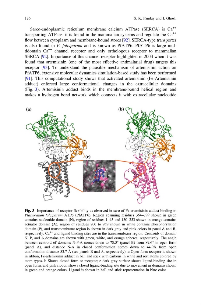
[91]. This computational study shows that activated artemisinin (Fe-Artemisinin
adduct) enforced large conformational changes in the extracellular domains
(Fig. 3). Artemisinin adduct binds in the membrane-bound helical region and
makes a hydrogen bond network which connects it with extracellular nucleotide
Fig. 3 Importance of receptor flexibility as observed in case of Fe-artemisinin adduct binding to
Plasmodium falciparum ATP6 (PfATP6). Region spanning residues 364–799 shown in green
contains nucleotide domain (N), region of residues 1–45 and 130–253 shown in orange contains
actuator domain (A), region of residues 800 to 959 shown in white contains phosphorylation
domain (P), and transmembrane region is shown in dark gray and pink colors in panel A and B,
respectively. Ca
++ and ligand binding sites are in the transmembrane region. Centroids of domain
N, P, and A domains are shown with green, white, and orange spheres, respectively. The angle
between centroid of domains N-P-A comes down to 78.5° (panel B) from 89.6° in open form
(panel A), and distance N-A in closed conformation comes down to 44.9Å from open
conformation distance 53.7 Å (see panels B and A, respectively). a Open-form receptor is shown
in ribbon, Fe-artemisinin adduct in ball and stick with carbons in white and rest atoms colored by
atom types. b Shows closed form or receptor; c dark gray surface shows ligand-binding site in
open form, and pink ribbon shows closed ligand-binding site due to movement in domains shown
in green and orange colors. Ligand is shown in ball and stick representation in blue color
126
S. K. Panday and I. Ghosh
++
transporting ATPase; it is found in the mammalian systems and regulate the Ca
++
flow between cytoplasm and membrane-bound stores [92]. SERCA-type transporter
is also found in P. falciparum and is known as PfATP6. PfATP6 is large multidomain Ca
++ channel receptor and only orthologous receptor to mammalian
SERCA [92]. Importance of this channel receptor highlighted in 2003 when it was
found that artemisinin (one of the most effective antimalarial drug) targets this
receptor [93]. To understand the plausible mechanism of artemisinin action on
PfATP6, extensive molecular dynamics simulation-based study has been performed
[91]. This computational study shows that activated artemisinin (Fe-Artemisinin
adduct) enforced large conformational changes in the extracellular domains
(Fig. 3). Artemisinin adduct binds in the membrane-bound helical region and
makes a hydrogen bond network which connects it with extracellular nucleotide
Fig. 3 Importance of receptor flexibility as observed in case of Fe-artemisinin adduct binding to
Plasmodium falciparum ATP6 (PfATP6). Region spanning residues 364–799 shown in green
contains nucleotide domain (N), region of residues 1–45 and 130–253 shown in orange contains
actuator domain (A), region of residues 800 to 959 shown in white contains phosphorylation
domain (P), and transmembrane region is shown in dark gray and pink colors in panel A and B,
respectively. Ca
++ and ligand binding sites are in the transmembrane region. Centroids of domain
N, P, and A domains are shown with green, white, and orange spheres, respectively. The angle
between centroid of domains N-P-A comes down to 78.5° (panel B) from 89.6° in open form
(panel A), and distance N-A in closed conformation comes down to 44.9Å from open
conformation distance 53.7 Å (see panels B and A, respectively). a Open-form receptor is shown
in ribbon, Fe-artemisinin adduct in ball and stick with carbons in white and rest atoms colored by
atom types. b Shows closed form or receptor; c dark gray surface shows ligand-binding site in
open form, and pink ribbon shows closed ligand-binding site due to movement in domains shown
in green and orange colors. Ligand is shown in ball and stick representation in blue color
126
S. K. Panday and I. Ghosh