

206
S. Sakkiah et al.
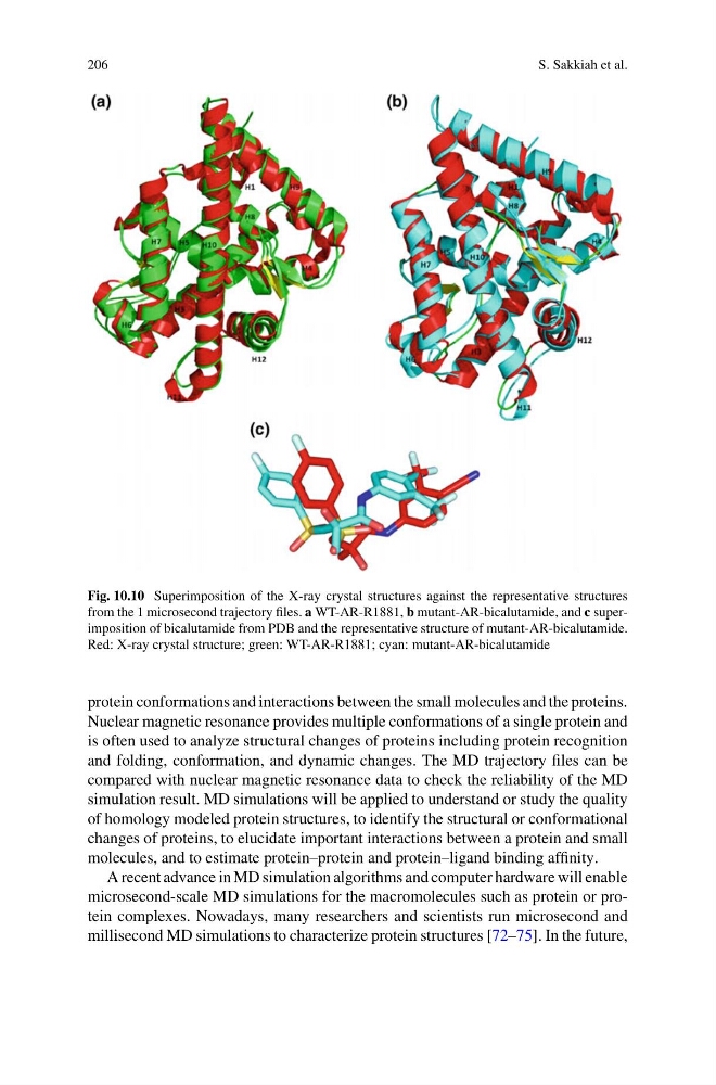
Fig. 10.10 Superimposition of the X-ray crystal structures against the representative structures
from the 1 microsecond trajectory files. a WT-AR-R1881, b mutant-AR-bicalutamide, and c superimposition of bicalutamide from PDB and the representative structure of mutant-AR-bicalutamide.
Red: X-ray crystal structure; green: WT-AR-R1881; cyan: mutant-AR-bicalutamide
protein conformations and interactions between the small molecules and the proteins.
Nuclear magnetic resonance provides multiple conformations of a single protein and
is often used to analyze structural changes of proteins including protein recognition
and folding, conformation, and dynamic changes. The MD trajectory files can be
compared with nuclear magnetic resonance data to check the reliability of the MD
simulation result. MD simulations will be applied to understand or study the quality
of homology modeled protein structures, to identify the structural or conformational
changes of proteins, to elucidate important interactions between a protein and small
molecules, and to estimate protein–protein and protein–ligand binding affinity.
A recent advance in MD simulation algorithms and computer hardware will enable
microsecond-scale MD simulations for the macromolecules such as protein or protein complexes. Nowadays, many researchers and scientists run microsecond and
millisecond MD simulations to characterize protein structures [72–75]. In the future,
S. Sakkiah et al.
Fig. 10.10 Superimposition of the X-ray crystal structures against the representative structures
from the 1 microsecond trajectory files. a WT-AR-R1881, b mutant-AR-bicalutamide, and c superimposition of bicalutamide from PDB and the representative structure of mutant-AR-bicalutamide.
Red: X-ray crystal structure; green: WT-AR-R1881; cyan: mutant-AR-bicalutamide
protein conformations and interactions between the small molecules and the proteins.
Nuclear magnetic resonance provides multiple conformations of a single protein and
is often used to analyze structural changes of proteins including protein recognition
and folding, conformation, and dynamic changes. The MD trajectory files can be
compared with nuclear magnetic resonance data to check the reliability of the MD
simulation result. MD simulations will be applied to understand or study the quality
of homology modeled protein structures, to identify the structural or conformational
changes of proteins, to elucidate important interactions between a protein and small
molecules, and to estimate protein–protein and protein–ligand binding affinity.
A recent advance in MD simulation algorithms and computer hardware will enable
microsecond-scale MD simulations for the macromolecules such as protein or protein complexes. Nowadays, many researchers and scientists run microsecond and
millisecond MD simulations to characterize protein structures [72–75]. In the future,