

8. Wash slides two times in PBS for 5 min and fix in fixation
solution for 24 h in darkness.
9. Perform sequential dehydration in 70, 90, and 100% ethanol
for 2 min each and leave to dry for 30 min.
10. Afterwards, perform pre-hybridization at 37
C for 1 night by
adding 15 μL FISH hybridization solution and cover slides
with coverslip. Store slides in a high-humidity plastic box.
11. Wash slides twice in 2Â SSC for 5 min and perform sequential
dehydration in 70, 90, and 100% ethanol for 2 min each. Leave
slides to dry for 30 min.
12. Perform DNA denaturation in denaturation solution at room
temperature for 10 min followed by sequential dehydration in
70, 90, and 100% ethanol for 2 min each. Leave slides to dry
for 15 min.
13. Meanwhile, prepare the FISH hybridization solution by mixing 0.5 μL of the 5
0 -labeled oligonucleotide probe (10 μM)
with 14.5 μL of FISH hybridization solution per slide.
14. Incubate the mixture at 95
C for 5 min followed by rapid
transfer onto ice for 5 min.
15. Apply 15 μL of the mixture per slide and hybridize at 37
C for
1 night. Cover slides with coverslips and store in a highhumidity plastic box.
16. Wash slides two times in 2Â SSC for 5 min followed by
sequential dehydration in 70, 90, and 100% ethanol for 2 min
each. Leave slides to dry for 30 min.
17. Apply 10 μL VECTASHIELD solution containing DAPI
(1:1000) and analyze slides by fluorescence microscopy
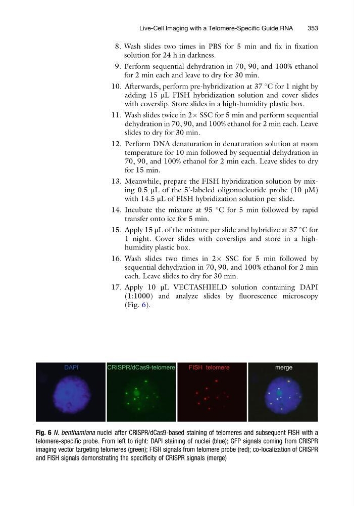
(Fig. 6).
Fig. 6 N. benthamiana nuclei after CRISPR/dCas9-based staining of telomeres and subsequent FISH with a
telomere-specific probe. From left to right: DAPI staining of nuclei (blue); GFP signals coming from CRISPR
imaging vector targeting telomeres (green); FISH signals from telomere probe (red); co-localization of CRISPR
and FISH signals demonstrating the specificity of CRISPR signals (merge)
Live-Cell Imaging with a Telomere-Specific Guide RNA
353
solution for 24 h in darkness.
9. Perform sequential dehydration in 70, 90, and 100% ethanol
for 2 min each and leave to dry for 30 min.
10. Afterwards, perform pre-hybridization at 37
C for 1 night by
adding 15 μL FISH hybridization solution and cover slides
with coverslip. Store slides in a high-humidity plastic box.
11. Wash slides twice in 2Â SSC for 5 min and perform sequential
dehydration in 70, 90, and 100% ethanol for 2 min each. Leave
slides to dry for 30 min.
12. Perform DNA denaturation in denaturation solution at room
temperature for 10 min followed by sequential dehydration in
70, 90, and 100% ethanol for 2 min each. Leave slides to dry
for 15 min.
13. Meanwhile, prepare the FISH hybridization solution by mixing 0.5 μL of the 5
0 -labeled oligonucleotide probe (10 μM)
with 14.5 μL of FISH hybridization solution per slide.
14. Incubate the mixture at 95
C for 5 min followed by rapid
transfer onto ice for 5 min.
15. Apply 15 μL of the mixture per slide and hybridize at 37
C for
1 night. Cover slides with coverslips and store in a highhumidity plastic box.
16. Wash slides two times in 2Â SSC for 5 min followed by
sequential dehydration in 70, 90, and 100% ethanol for 2 min
each. Leave slides to dry for 30 min.
17. Apply 10 μL VECTASHIELD solution containing DAPI
(1:1000) and analyze slides by fluorescence microscopy
(Fig. 6).
Fig. 6 N. benthamiana nuclei after CRISPR/dCas9-based staining of telomeres and subsequent FISH with a
telomere-specific probe. From left to right: DAPI staining of nuclei (blue); GFP signals coming from CRISPR
imaging vector targeting telomeres (green); FISH signals from telomere probe (red); co-localization of CRISPR
and FISH signals demonstrating the specificity of CRISPR signals (merge)
Live-Cell Imaging with a Telomere-Specific Guide RNA
353