

photobleaching of SERS labels and autofluorescence of samples as well as narrow
SERS signals compared to fluorescence that permits multiplexing capabilities
[191].
iSERS imaging of prostate tissues to detect prostate-specific antigen (PSA) was
proposed firstly by Schlücker et al. in 2006 [192]. Next, they extended this
methodology for monitoring PSA expression [193] and determining co-localization
of p63 and PSA on healthy prostate tissue [194]. The latter was the first report
showing multiplexing capability of iSERS in tissues; for this purpose, two different
Raman reporters: 4-nitrothiobenzoic acid (4-NTB) and 4-mercaptobenzoic acid
(4-MBA) were employed to construct iSERS nanotags. The Schlücker group also
demonstrated a successful iSERS-based localization and expression of the HER2 in
normal and malignant breast tissues [195]. A similar approach was used by Wang
and co-workers for quantitative multiplexed imaging of freshly excised human
tissues for surgical guidance applications [196]. Ex vivo experiments carried out by
Quynh et al. [197] confirmed the utility of iSERS imaging to detect tumor regions.
Here, BerEP4 antibody conjugated to SERS NPs covered by 4-ATP
(4-aminothiophenol) as a Raman reporter specifically recognized basal cell carcinoma (BCC) in skin during surgical procedures. SERS detection of antibody–
antigen interactions was also used for a rapid and sensitive recognition of LMP1
protein expression specific for nasopharyngeal cancer [198].
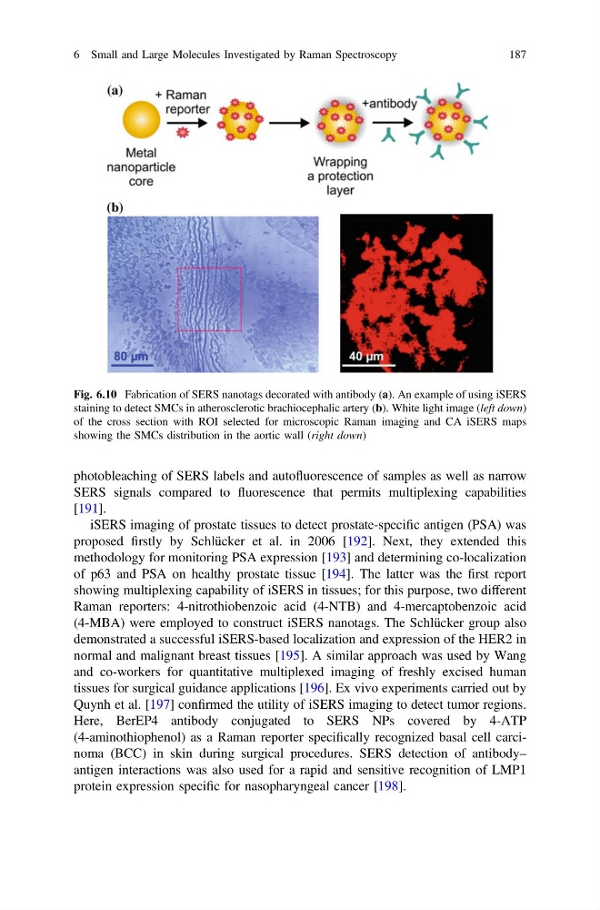
Fig. 6.10 Fabrication of SERS nanotags decorated with antibody (a). An example of using iSERS
staining to detect SMCs in atherosclerotic brachiocephalic artery (b). White light image (left down)
of the cross section with ROI selected for microscopic Raman imaging and CA iSERS maps
showing the SMCs distribution in the aortic wall (right down)
6 Small and Large Molecules Investigated by Raman Spectroscopy
187
SERS signals compared to fluorescence that permits multiplexing capabilities
[191].
iSERS imaging of prostate tissues to detect prostate-specific antigen (PSA) was
proposed firstly by Schlücker et al. in 2006 [192]. Next, they extended this
methodology for monitoring PSA expression [193] and determining co-localization
of p63 and PSA on healthy prostate tissue [194]. The latter was the first report
showing multiplexing capability of iSERS in tissues; for this purpose, two different
Raman reporters: 4-nitrothiobenzoic acid (4-NTB) and 4-mercaptobenzoic acid
(4-MBA) were employed to construct iSERS nanotags. The Schlücker group also
demonstrated a successful iSERS-based localization and expression of the HER2 in
normal and malignant breast tissues [195]. A similar approach was used by Wang
and co-workers for quantitative multiplexed imaging of freshly excised human
tissues for surgical guidance applications [196]. Ex vivo experiments carried out by
Quynh et al. [197] confirmed the utility of iSERS imaging to detect tumor regions.
Here, BerEP4 antibody conjugated to SERS NPs covered by 4-ATP
(4-aminothiophenol) as a Raman reporter specifically recognized basal cell carcinoma (BCC) in skin during surgical procedures. SERS detection of antibody–
antigen interactions was also used for a rapid and sensitive recognition of LMP1
protein expression specific for nasopharyngeal cancer [198].
Fig. 6.10 Fabrication of SERS nanotags decorated with antibody (a). An example of using iSERS
staining to detect SMCs in atherosclerotic brachiocephalic artery (b). White light image (left down)
of the cross section with ROI selected for microscopic Raman imaging and CA iSERS maps
showing the SMCs distribution in the aortic wall (right down)
6 Small and Large Molecules Investigated by Raman Spectroscopy
187