

66
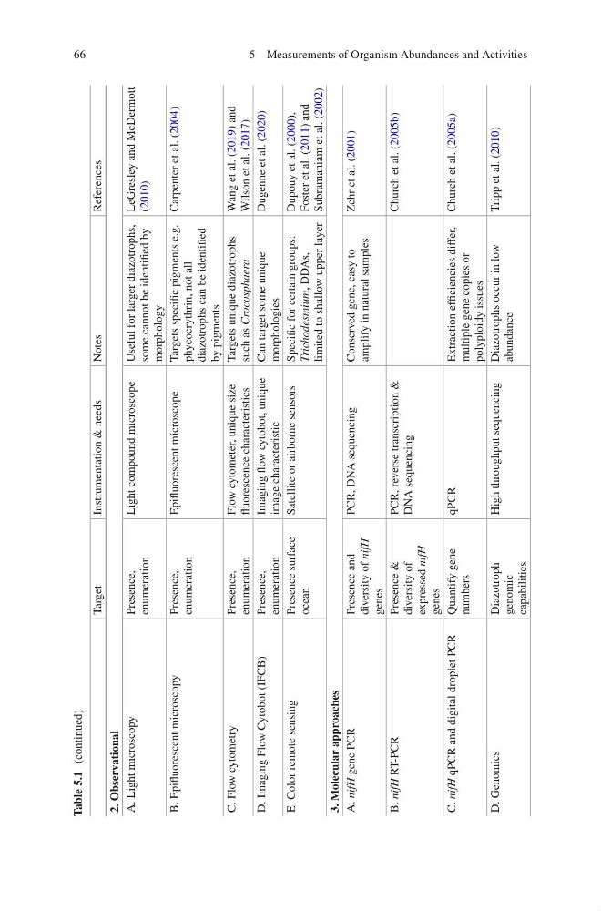
Table 5.1 (continued)
Target
Instrumentation & needs
Notes
References
2. Observational
A. Light microscopy
Presence,
enumeration
Light compound microscope
Useful for larger diazotrophs,
some cannot be identified by
morphology
LeGresley and McDermott
(2010)
B. Epifluorescent microscopy
Presence,
enumeration
Epifluorescent microscope
Targets specific pigments e.g.
phycoerythrin, not all
diazotrophs can be identified
by pigments
Carpenter et al. (2004)
C. Flow cytometry
Presence,
enumeration
Flow cytometer, unique size
fluorescence characteristics
Targets unique diazotrophs
such as Crocosphaera
Wang et al. (2019) and
Wilson et al. (2017)
D. Imaging Flow Cytobot (IFCB)
Presence,
enumeration
Imaging flow cytobot, unique
image characteristic
Can target some unique
morphologies
Dugenne et al. (2020)
E. Color remote sensing
Presence surface
ocean
Satellite or airborne sensors
Specific for certain groups:
Trichodesmium,
DDAs,
limited to shallow upper layer
Dupouy et al. (2000),
Foster et al. (2011) and
Subramaniam et al. (2002)
3. Molecular approaches
A. nifH
gene PCR
Presence and
diversity of nifH
genes
PCR, DNA sequencing
Conserved gene, easy to
amplify in natural samples
Zehr et al. (2001)
B. nifH
RT-PCR
Presence &
diversity of
expressed nifH
genes
PCR, reverse transcription &
DNA sequencing
Church et al. (2005b)
C. nifH
qPCR and digital droplet PCR
Quantify gene
numbers
qPCR
Extraction efficiencies differ,
multiple gene copies or
polyploidy issues
Church et al. (2005a)
D. Genomics
Diazotroph
genomic
capabilities
High throughput sequencing
Diazotrophs occur in low
abundance
Tripp et al. (2010)
5 Measurements of Organism Abundances and Activities
Table 5.1 (continued)
Target
Instrumentation & needs
Notes
References
2. Observational
A. Light microscopy
Presence,
enumeration
Light compound microscope
Useful for larger diazotrophs,
some cannot be identified by
morphology
LeGresley and McDermott
(2010)
B. Epifluorescent microscopy
Presence,
enumeration
Epifluorescent microscope
Targets specific pigments e.g.
phycoerythrin, not all
diazotrophs can be identified
by pigments
Carpenter et al. (2004)
C. Flow cytometry
Presence,
enumeration
Flow cytometer, unique size
fluorescence characteristics
Targets unique diazotrophs
such as Crocosphaera
Wang et al. (2019) and
Wilson et al. (2017)
D. Imaging Flow Cytobot (IFCB)
Presence,
enumeration
Imaging flow cytobot, unique
image characteristic
Can target some unique
morphologies
Dugenne et al. (2020)
E. Color remote sensing
Presence surface
ocean
Satellite or airborne sensors
Specific for certain groups:
Trichodesmium,
DDAs,
limited to shallow upper layer
Dupouy et al. (2000),
Foster et al. (2011) and
Subramaniam et al. (2002)
3. Molecular approaches
A. nifH
gene PCR
Presence and
diversity of nifH
genes
PCR, DNA sequencing
Conserved gene, easy to
amplify in natural samples
Zehr et al. (2001)
B. nifH
RT-PCR
Presence &
diversity of
expressed nifH
genes
PCR, reverse transcription &
DNA sequencing
Church et al. (2005b)
C. nifH
qPCR and digital droplet PCR
Quantify gene
numbers
qPCR
Extraction efficiencies differ,
multiple gene copies or
polyploidy issues
Church et al. (2005a)
D. Genomics
Diazotroph
genomic
capabilities
High throughput sequencing
Diazotrophs occur in low
abundance
Tripp et al. (2010)
5 Measurements of Organism Abundances and Activities