

52
T. B. Asafa et al.
2.8 Magnetic Force Microscopy
The principle of magnetic force microscopy (MFM) is quite similar to that of the
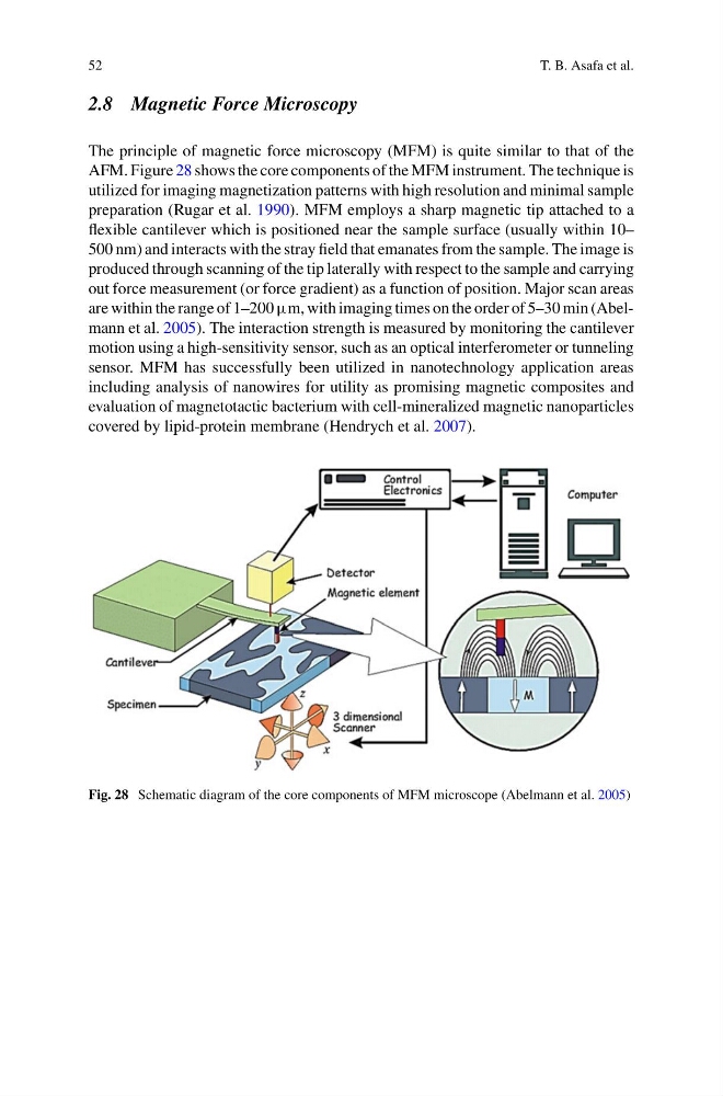
AFM. Figure 28 shows the core components of the MFM instrument. The technique is
utilized for imaging magnetization patterns with high resolution and minimal sample
preparation (Rugar et al. 1990). MFM employs a sharp magnetic tip attached to a
flexible cantilever which is positioned near the sample surface (usually within 10–
500 nm) and interacts with the stray field that emanates from the sample. The image is
produced through scanning of the tip laterally with respect to the sample and carrying
out force measurement (or force gradient) as a function of position. Major scan areas
are within the range of 1–200 μm, with imaging times on the order of 5–30 min (Abelmann et al. 2005). The interaction strength is measured by monitoring the cantilever
motion using a high-sensitivity sensor, such as an optical interferometer or tunneling
sensor. MFM has successfully been utilized in nanotechnology application areas
including analysis of nanowires for utility as promising magnetic composites and
evaluation of magnetotactic bacterium with cell-mineralized magnetic nanoparticles
covered by lipid-protein membrane (Hendrych et al. 2007).
Fig. 28 Schematic diagram of the core components of MFM microscope (Abelmann et al. 2005)
T. B. Asafa et al.
2.8 Magnetic Force Microscopy
The principle of magnetic force microscopy (MFM) is quite similar to that of the
AFM. Figure 28 shows the core components of the MFM instrument. The technique is
utilized for imaging magnetization patterns with high resolution and minimal sample
preparation (Rugar et al. 1990). MFM employs a sharp magnetic tip attached to a
flexible cantilever which is positioned near the sample surface (usually within 10–
500 nm) and interacts with the stray field that emanates from the sample. The image is
produced through scanning of the tip laterally with respect to the sample and carrying
out force measurement (or force gradient) as a function of position. Major scan areas
are within the range of 1–200 μm, with imaging times on the order of 5–30 min (Abelmann et al. 2005). The interaction strength is measured by monitoring the cantilever
motion using a high-sensitivity sensor, such as an optical interferometer or tunneling
sensor. MFM has successfully been utilized in nanotechnology application areas
including analysis of nanowires for utility as promising magnetic composites and
evaluation of magnetotactic bacterium with cell-mineralized magnetic nanoparticles
covered by lipid-protein membrane (Hendrych et al. 2007).
Fig. 28 Schematic diagram of the core components of MFM microscope (Abelmann et al. 2005)