

Characterization Techniques in Nanotechnology …
41
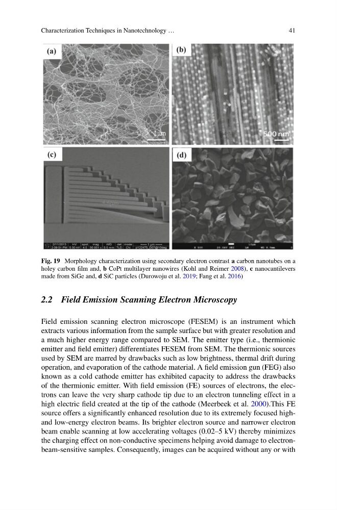
Fig. 19 Morphology characterization using secondary electron contrast a carbon nanotubes on a
holey carbon film and, b CoPt multilayer nanowires (Kohl and Reimer 2008), c nanocantilevers
made from SiGe and, d SiC particles (Durowoju et al. 2019; Fang et al. 2016)
2.2 Field Emission Scanning Electron Microscopy
Field emission scanning electron microscope (FESEM) is an instrument which
extracts various information from the sample surface but with greater resolution and
a much higher energy range compared to SEM. The emitter type (i.e., thermionic
emitter and field emitter) differentiates FESEM from SEM. The thermionic sources
used by SEM are marred by drawbacks such as low brightness, thermal drift during
operation, and evaporation of the cathode material. A field emission gun (FEG) also
known as a cold cathode emitter has exhibited capacity to address the drawbacks
of the thermionic emitter. With field emission (FE) sources of electrons, the electrons can leave the very sharp cathode tip due to an electron tunneling effect in a
high electric field created at the tip of the cathode (Meerbeek et al. 2000).This FE
source offers a significantly enhanced resolution due to its extremely focused highand low-energy electron beams. Its brighter electron source and narrower electron
beam enable scanning at low accelerating voltages (0.02–5 kV) thereby minimizes
the charging effect on non-conductive specimens helping avoid damage to electronbeam-sensitive samples. Consequently, images can be acquired without any or with
41
Fig. 19 Morphology characterization using secondary electron contrast a carbon nanotubes on a
holey carbon film and, b CoPt multilayer nanowires (Kohl and Reimer 2008), c nanocantilevers
made from SiGe and, d SiC particles (Durowoju et al. 2019; Fang et al. 2016)
2.2 Field Emission Scanning Electron Microscopy
Field emission scanning electron microscope (FESEM) is an instrument which
extracts various information from the sample surface but with greater resolution and
a much higher energy range compared to SEM. The emitter type (i.e., thermionic
emitter and field emitter) differentiates FESEM from SEM. The thermionic sources
used by SEM are marred by drawbacks such as low brightness, thermal drift during
operation, and evaporation of the cathode material. A field emission gun (FEG) also
known as a cold cathode emitter has exhibited capacity to address the drawbacks
of the thermionic emitter. With field emission (FE) sources of electrons, the electrons can leave the very sharp cathode tip due to an electron tunneling effect in a
high electric field created at the tip of the cathode (Meerbeek et al. 2000).This FE
source offers a significantly enhanced resolution due to its extremely focused highand low-energy electron beams. Its brighter electron source and narrower electron
beam enable scanning at low accelerating voltages (0.02–5 kV) thereby minimizes
the charging effect on non-conductive specimens helping avoid damage to electronbeam-sensitive samples. Consequently, images can be acquired without any or with