

Shen et al. (2018) prepared carbon quantum dots coupled with TiO 2 via hydrothermal method using glucose and citric acid as precursors. The heterojunction
exhibits enhanced photocatalytic activity against phenol under ultraviolet light.
Carbon quantum dots–glucose/TiO 2 heterojunction was synthesized using glucose
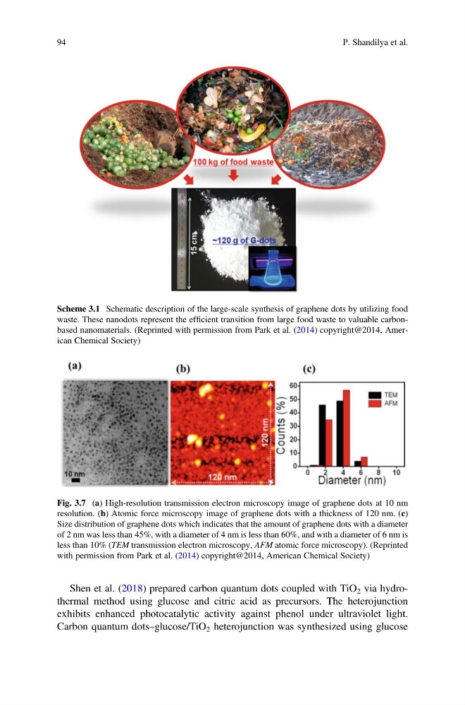
Scheme 3.1 Schematic description of the large-scale synthesis of graphene dots by utilizing food
waste. These nanodots represent the efficient transition from large food waste to valuable carbonbased nanomaterials. (Reprinted with permission from Park et al. (2014) copyright@2014, American Chemical Society)
Fig. 3.7 (a) High-resolution transmission electron microscopy image of graphene dots at 10 nm
resolution. (b) Atomic force microscopy image of graphene dots with a thickness of 120 nm. (c)
Size distribution of graphene dots which indicates that the amount of graphene dots with a diameter
of 2 nm was less than 45%, with a diameter of 4 nm is less than 60%, and with a diameter of 6 nm is
less than 10% (TEM transmission electron microscopy, AFM atomic force microscopy). (Reprinted
with permission from Park et al. (2014) copyright@2014, American Chemical Society)
94
P. Shandilya et al.
exhibits enhanced photocatalytic activity against phenol under ultraviolet light.
Carbon quantum dots–glucose/TiO 2 heterojunction was synthesized using glucose
Scheme 3.1 Schematic description of the large-scale synthesis of graphene dots by utilizing food
waste. These nanodots represent the efficient transition from large food waste to valuable carbonbased nanomaterials. (Reprinted with permission from Park et al. (2014) copyright@2014, American Chemical Society)
Fig. 3.7 (a) High-resolution transmission electron microscopy image of graphene dots at 10 nm
resolution. (b) Atomic force microscopy image of graphene dots with a thickness of 120 nm. (c)
Size distribution of graphene dots which indicates that the amount of graphene dots with a diameter
of 2 nm was less than 45%, with a diameter of 4 nm is less than 60%, and with a diameter of 6 nm is
less than 10% (TEM transmission electron microscopy, AFM atomic force microscopy). (Reprinted
with permission from Park et al. (2014) copyright@2014, American Chemical Society)
94
P. Shandilya et al.