

88
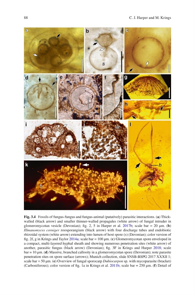
Fig. 3.4 Fossils of fungus-fungus and fungus-animal (putatively) parasitic interactions. (a) Thickwalled (black arrow) and smaller thinner-walled propagules (white arrow) of fungal intruder in
glomeromycotan vesicle (Devonian); fig. 2, 5 in Harper et al. 2017b; scale bar = 20 μm. (b)
Illmanomyces corniger zoosporangium (black arrow) with four discharge tubes and endobiotic
rhizoidal system (white arrow) extending into lumen of host spore (s) (Devonian); color version of
fig. 2f, g in Krings and Taylor 2014a; scale bar = 100 μm. (c) Glomeromycotan spore enveloped in
a compact, multi-layered hyphal sheath and showing numerous penetration sites (white arrow) of
another, parasitic fungus (black arrow) (Devonian); fig. 3F in Krings and Harper 2018; scale
bar = 10 μm. (d) Massive, branched callosity in a glomeromycotan spore (Devonian); note parasite
penetration sites on spore surface (arrows); Munich collection, slide SNSB-BSPG 2017 XXXII 1;
scale bar = 50 μm. (e) Overview of fungal sporocarp Dubiocarpon sp. with mycoparasite (bracket)
(Carboniferous); color version of fig. 1a in Krings et al. 2011b; scale bar = 250 μm. (f) Detail of
C. J. Harper and M. Krings
Fig. 3.4 Fossils of fungus-fungus and fungus-animal (putatively) parasitic interactions. (a) Thickwalled (black arrow) and smaller thinner-walled propagules (white arrow) of fungal intruder in
glomeromycotan vesicle (Devonian); fig. 2, 5 in Harper et al. 2017b; scale bar = 20 μm. (b)
Illmanomyces corniger zoosporangium (black arrow) with four discharge tubes and endobiotic
rhizoidal system (white arrow) extending into lumen of host spore (s) (Devonian); color version of
fig. 2f, g in Krings and Taylor 2014a; scale bar = 100 μm. (c) Glomeromycotan spore enveloped in
a compact, multi-layered hyphal sheath and showing numerous penetration sites (white arrow) of
another, parasitic fungus (black arrow) (Devonian); fig. 3F in Krings and Harper 2018; scale
bar = 10 μm. (d) Massive, branched callosity in a glomeromycotan spore (Devonian); note parasite
penetration sites on spore surface (arrows); Munich collection, slide SNSB-BSPG 2017 XXXII 1;
scale bar = 50 μm. (e) Overview of fungal sporocarp Dubiocarpon sp. with mycoparasite (bracket)
(Carboniferous); color version of fig. 1a in Krings et al. 2011b; scale bar = 250 μm. (f) Detail of
C. J. Harper and M. Krings