

32
from 0.2 to 3.0 μm in diameter. In some, putative isometric virions are barely visible
as dark specks (Fig. 2.4). Extant cypoviruses infect the midgut of a wide range of
insects, including Ceratopogonidae, along with a variety of other lower Diptera
(Nematocera) (Poinar and Thomas 1984; Poinar and Poinar 2005).
Also in Myanmar amber is evidence of a nuclear polyhedrosis virus (NPV)
(Baculoviridae) infecting a sand fly (Diptera: Phlebotomidae) (Poinar and Poinar
2005) (Figs. 2.5 and 2.6). The inclusion bodies (polyhedra) are irregular and in
some, putative rod-shaped virions are barely visible as dark rods (Fig. 2.7). Nuclear
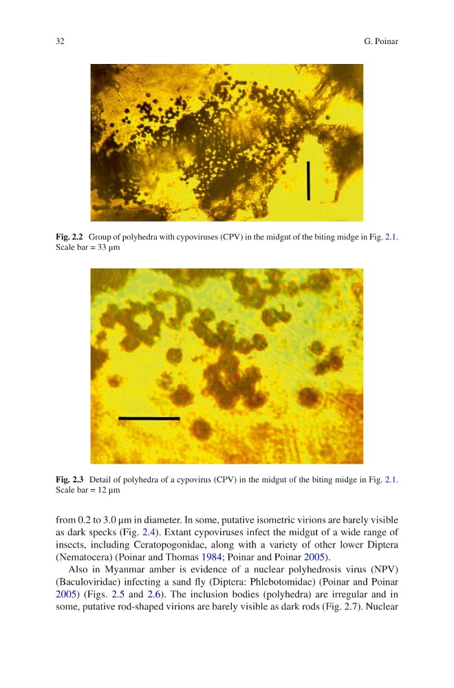
Fig. 2.2 Group of polyhedra with cypoviruses (CPV) in the midgut of the biting midge in Fig. 2.1.
Scale bar = 33 μm
Fig. 2.3 Detail of polyhedra of a cypovirus (CPV) in the midgut of the biting midge in Fig. 2.1.
Scale bar = 12 μm
G. Poinar
from 0.2 to 3.0 μm in diameter. In some, putative isometric virions are barely visible
as dark specks (Fig. 2.4). Extant cypoviruses infect the midgut of a wide range of
insects, including Ceratopogonidae, along with a variety of other lower Diptera
(Nematocera) (Poinar and Thomas 1984; Poinar and Poinar 2005).
Also in Myanmar amber is evidence of a nuclear polyhedrosis virus (NPV)
(Baculoviridae) infecting a sand fly (Diptera: Phlebotomidae) (Poinar and Poinar
2005) (Figs. 2.5 and 2.6). The inclusion bodies (polyhedra) are irregular and in
some, putative rod-shaped virions are barely visible as dark rods (Fig. 2.7). Nuclear
Fig. 2.2 Group of polyhedra with cypoviruses (CPV) in the midgut of the biting midge in Fig. 2.1.
Scale bar = 33 μm
Fig. 2.3 Detail of polyhedra of a cypovirus (CPV) in the midgut of the biting midge in Fig. 2.1.
Scale bar = 12 μm
G. Poinar