

31
Indirect evidence of virus infections is when some modification of an organism,
such as color or form that has been shown to be the result of an extant virus infection
is recovered in the fossil state. Also included in this category are fossil organisms
whose present day descendants are known to harbor genomic viruses such as
polydnaviruses.
2.2.1 Direct Evidence of Fossil Viral Infections
Several groups of insect pathogenic viruses are known as occluded viruses since
their virions are embedded in a crystaline protein lattice called a polyhedron or
inclusion body. Some insect polyhedra are large enough to be seen with the light
microscope and their identification can be based on their shape, size, location and
dimensions of the embedded virions. Two viral groups that form relatively large
polyhedra visible under the light microscope (Poinar and Thomas 1984) are the
cypoviruses (cytoplasmic polyhedrosis viruses or CPV such as Reoviridae) and the
nuclear polyhedrosis viruses (NPV such as Baculoviridae). Fossil examples of both
of these viral types have been identified in Myanmar amber (Poinar and Poinar
2005), dated to the mid-Cretaceous (Cruickhank and Ko 2003; Shi et al. 2012).
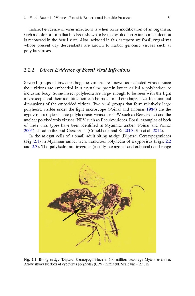
In the midgut cells of a small adult biting midge (Diptera; Ceratopogonidae)
(Fig. 2.1) in Myanmar amber were numerous polyhedra of a cypovirus (Figs. 2.2
and 2.3). The polyhedra are irregular (mostly hexagonal and cuboidal) and range
Fig. 2.1 Biting midge (Diptera: Ceratopogonidae) in 100 million years ago Myanmar amber.
Arrow shows location of cypovirus polyhedra (CPV) in midgut. Scale bar = 22 μm
2 Fossil Record of Viruses, Parasitic Bacteria and Parasitic Protozoa
Indirect evidence of virus infections is when some modification of an organism,
such as color or form that has been shown to be the result of an extant virus infection
is recovered in the fossil state. Also included in this category are fossil organisms
whose present day descendants are known to harbor genomic viruses such as
polydnaviruses.
2.2.1 Direct Evidence of Fossil Viral Infections
Several groups of insect pathogenic viruses are known as occluded viruses since
their virions are embedded in a crystaline protein lattice called a polyhedron or
inclusion body. Some insect polyhedra are large enough to be seen with the light
microscope and their identification can be based on their shape, size, location and
dimensions of the embedded virions. Two viral groups that form relatively large
polyhedra visible under the light microscope (Poinar and Thomas 1984) are the
cypoviruses (cytoplasmic polyhedrosis viruses or CPV such as Reoviridae) and the
nuclear polyhedrosis viruses (NPV such as Baculoviridae). Fossil examples of both
of these viral types have been identified in Myanmar amber (Poinar and Poinar
2005), dated to the mid-Cretaceous (Cruickhank and Ko 2003; Shi et al. 2012).
In the midgut cells of a small adult biting midge (Diptera; Ceratopogonidae)
(Fig. 2.1) in Myanmar amber were numerous polyhedra of a cypovirus (Figs. 2.2
and 2.3). The polyhedra are irregular (mostly hexagonal and cuboidal) and range
Fig. 2.1 Biting midge (Diptera: Ceratopogonidae) in 100 million years ago Myanmar amber.
Arrow shows location of cypovirus polyhedra (CPV) in midgut. Scale bar = 22 μm
2 Fossil Record of Viruses, Parasitic Bacteria and Parasitic Protozoa