

Topics in Current Chemistry (2020) 378:40
1 3
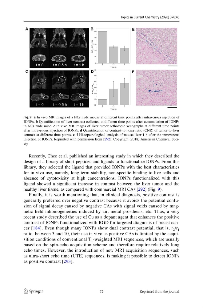
Recently, Chee et al. published an interesting study in which they described the
design of a library of short peptides and ligands to functionalize IONPs. From this
library, they selected the ligand that provided IONPs with the best characteristics
for in vivo use, namely, long term stability, non-specific binding to live cells and
absence of cytotoxicity at high concentrations. IONPs functionalized with this
ligand showed a significant increase in contrast between the liver tumor and the
healthy liver tissue, as compared with commercial MRI CAs [292] (Fig. 9).
Finally, it is worth mentioning that, in clinical diagnosis, positive contrast is
generally preferred over negative contrast because it avoids the potential confusion of signal decay caused by negative CAs with signal voids caused by magnetic field inhomogeneities induced by air, metal prosthesis, etc. Thus, a very
recent study described the use of Cu as a dopant agent that enhances the positive
contrast of IONPs functionalized with RGD for targeted diagnosis of breast cancer [184]. Even though many IONPs show dual contrast potential, that is, r 2 /r 1
ratio between 3 and 10, their use in vivo as positive CAs is limited by the acquisition conditions of conventional T 1 -weighted MRI sequences, which are usually
based on the spin-echo acquisition scheme and therefore require relatively long
echo times. However, the introduction of new MRI acquisition sequences, such
as ultra-short echo time (UTE) sequences, is making it possible to detect IONPs
as positive contrast [293].
Fig. 9 a In vivo MR images of a NCr nude mouse at different time points after intravenous injection of
IONPs. b Quantification of liver contrast collected at different time points after accumulation of IONPs
in NCr nude mice. c In vivo MR images of liver tumor orthotopic xenographs at different time points
after intravenous injection of IONPs. d Quantification of contrast-to-noise ratio (CNR) of tumor-to-liver
contrast at different time points. e, f Histopathological analysis of mouse liver 1 h after the intravenous
injection of IONPs. Reprinted with permission from [292]. Copyright (2018) American Chemical Society
72
Reprinted from the journal
1 3
Recently, Chee et al. published an interesting study in which they described the
design of a library of short peptides and ligands to functionalize IONPs. From this
library, they selected the ligand that provided IONPs with the best characteristics
for in vivo use, namely, long term stability, non-specific binding to live cells and
absence of cytotoxicity at high concentrations. IONPs functionalized with this
ligand showed a significant increase in contrast between the liver tumor and the
healthy liver tissue, as compared with commercial MRI CAs [292] (Fig. 9).
Finally, it is worth mentioning that, in clinical diagnosis, positive contrast is
generally preferred over negative contrast because it avoids the potential confusion of signal decay caused by negative CAs with signal voids caused by magnetic field inhomogeneities induced by air, metal prosthesis, etc. Thus, a very
recent study described the use of Cu as a dopant agent that enhances the positive
contrast of IONPs functionalized with RGD for targeted diagnosis of breast cancer [184]. Even though many IONPs show dual contrast potential, that is, r 2 /r 1
ratio between 3 and 10, their use in vivo as positive CAs is limited by the acquisition conditions of conventional T 1 -weighted MRI sequences, which are usually
based on the spin-echo acquisition scheme and therefore require relatively long
echo times. However, the introduction of new MRI acquisition sequences, such
as ultra-short echo time (UTE) sequences, is making it possible to detect IONPs
as positive contrast [293].
Fig. 9 a In vivo MR images of a NCr nude mouse at different time points after intravenous injection of
IONPs. b Quantification of liver contrast collected at different time points after accumulation of IONPs
in NCr nude mice. c In vivo MR images of liver tumor orthotopic xenographs at different time points
after intravenous injection of IONPs. d Quantification of contrast-to-noise ratio (CNR) of tumor-to-liver
contrast at different time points. e, f Histopathological analysis of mouse liver 1 h after the intravenous
injection of IONPs. Reprinted with permission from [292]. Copyright (2018) American Chemical Society
72
Reprinted from the journal