

Topics in Current Chemistry (2020) 378:40
1 3
3.3 Ligand Exchange
Ligand exchange is a very complicated coating strategy that involves multiple interactions potencials/forces. It requires the use of reactive binding molecules that enable the replacement of capping agents attached to the nanoparticle surfaces. This
binding between the iron atoms of the IONP and the anchor group of the ligand molecules is mediated by electrostatic interactions. Therefore, the nature of the anchor
group is determinant in the search for highly stable ligand molecules at the IONP
surfaces. In addition to anchor groups, the hydrophilic balance of the ligand is also
important to render water-soluble NPs [236, 246, 247]. In our group, we have developed different ligand formulations to functionalize IONPs to obtain soluble and
stable NPs in physiological media for in vivo MRI applications. These ligands are
based on a gallol group as a strong binder and PEG chains as hydrophilic tunable
spacers, which also minimize plasma protein adsorption. In this manner, we have
demonstrated that selection of the right molecular weight of PEG chain and the outermost charged group of the ligand plays a fundamental role in the fate and bioavailability of intravenously injected IONPs. Thus, a ligand with a PEG chain between
1500 and 3000 Da and neutral outermost groups showed the best stealth properties, resulting in longer blood circulation times and higher bioavailability without
increased toxicity [248–250].
4 Applications of IONPs in MRI
Among the main clinical diagnostic techniques, MRI stands out for its unique combination of qualities, such as its non-invasive character, the absence of ionizing
radiation, excellent image quality, and its ability to provide both anatomical and
functional information [251]. The MRI signal comes mainly from the protons of
the water molecules, while the image contrast is generated from differences in the
intensity of this signal among different tissues, which depends on the concentration,
relaxation times (T 1 and T 2 ) and mobility of the water molecules within each tissue [252, 253]. Additionally, image contrast can be further enhanced using CAs.
Although there are several mechanisms that can produce MRI contrast, such as
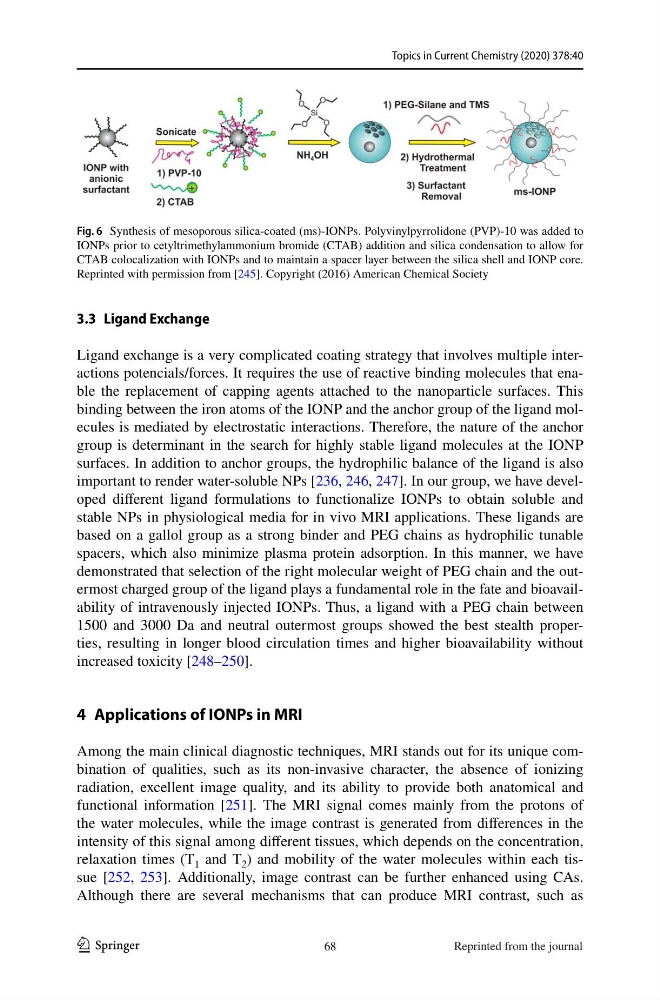
Fig. 6 Synthesis of mesoporous silica-coated (ms)-IONPs. Polyvinylpyrrolidone (PVP)-10 was added to
IONPs prior to cetyltrimethylammonium bromide (CTAB) addition and silica condensation to allow for
CTAB colocalization with IONPs and to maintain a spacer layer between the silica shell and IONP core.
Reprinted with permission from [245]. Copyright (2016) American Chemical Society
68
Reprinted from the journal
1 3
3.3 Ligand Exchange
Ligand exchange is a very complicated coating strategy that involves multiple interactions potencials/forces. It requires the use of reactive binding molecules that enable the replacement of capping agents attached to the nanoparticle surfaces. This
binding between the iron atoms of the IONP and the anchor group of the ligand molecules is mediated by electrostatic interactions. Therefore, the nature of the anchor
group is determinant in the search for highly stable ligand molecules at the IONP
surfaces. In addition to anchor groups, the hydrophilic balance of the ligand is also
important to render water-soluble NPs [236, 246, 247]. In our group, we have developed different ligand formulations to functionalize IONPs to obtain soluble and
stable NPs in physiological media for in vivo MRI applications. These ligands are
based on a gallol group as a strong binder and PEG chains as hydrophilic tunable
spacers, which also minimize plasma protein adsorption. In this manner, we have
demonstrated that selection of the right molecular weight of PEG chain and the outermost charged group of the ligand plays a fundamental role in the fate and bioavailability of intravenously injected IONPs. Thus, a ligand with a PEG chain between
1500 and 3000 Da and neutral outermost groups showed the best stealth properties, resulting in longer blood circulation times and higher bioavailability without
increased toxicity [248–250].
4 Applications of IONPs in MRI
Among the main clinical diagnostic techniques, MRI stands out for its unique combination of qualities, such as its non-invasive character, the absence of ionizing
radiation, excellent image quality, and its ability to provide both anatomical and
functional information [251]. The MRI signal comes mainly from the protons of
the water molecules, while the image contrast is generated from differences in the
intensity of this signal among different tissues, which depends on the concentration,
relaxation times (T 1 and T 2 ) and mobility of the water molecules within each tissue [252, 253]. Additionally, image contrast can be further enhanced using CAs.
Although there are several mechanisms that can produce MRI contrast, such as
Fig. 6 Synthesis of mesoporous silica-coated (ms)-IONPs. Polyvinylpyrrolidone (PVP)-10 was added to
IONPs prior to cetyltrimethylammonium bromide (CTAB) addition and silica condensation to allow for
CTAB colocalization with IONPs and to maintain a spacer layer between the silica shell and IONP core.
Reprinted with permission from [245]. Copyright (2016) American Chemical Society
68
Reprinted from the journal