

Topics in Current Chemistry (2020) 378:28
1 3
matrix was employed to anchor Ab 1 . Finally, insulin was sensitively measured
with MnO 2 @C nanospheres used as an energy-transfer quenching label.
Various applications of ECL microscopy appear in the literature due to the
extremely low background light and high throughput of this technique. Because of
the steric hindrance and low electrical conductivity of cells, it is difficult to perform
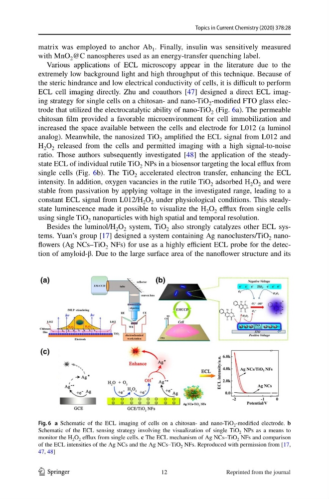
ECL cell imaging directly. Zhu and coauthors [47] designed a direct ECL imaging strategy for single cells on a chitosan- and nano-TiO 2 -modified FTO glass electrode that utilized the electrocatalytic ability of nano-TiO 2 (Fig. 6a). The permeable
chitosan film provided a favorable microenvironment for cell immobilization and
increased the space available between the cells and electrode for L012 (a luminol
analog). Meanwhile, the nanosized TiO 2 amplified the ECL signal from L012 and
H 2 O 2 released from the cells and permitted imaging with a high signal-to-noise
ratio. Those authors subsequently investigated [48] the application of the steadystate ECL of individual rutile TiO 2 NPs in a biosensor targeting the local efflux from
single cells (Fig. 6b). The TiO 2 accelerated electron transfer, enhancing the ECL
intensity. In addition, oxygen vacancies in the rutile TiO 2 adsorbed H 2 O 2 and were
stable from passivation by applying voltage in the investigated range, leading to a
constant ECL signal from L012/H 2 O 2 under physiological conditions. This steadystate luminescence made it possible to visualize the H 2 O 2 efflux from single cells
using single TiO 2 nanoparticles with high spatial and temporal resolution.
Besides the luminol/H 2 O 2 system, TiO 2 also strongly catalyzes other ECL systems. Yuan’s group [17] designed a system containing Ag nanoclusters/TiO 2 nanoflowers (Ag NCs–TiO 2 NFs) for use as a highly efficient ECL probe for the detection of amyloid-β. Due to the large surface area of the nanoflower structure and its
Fig. 6 a Schematic of the ECL imaging of cells on a chitosan- and nano-TiO 2 -modified electrode. b
Schematic of the ECL sensing strategy involving the visualization of single TiO 2 NPs as a means to
monitor the H 2 O 2 efflux from single cells. c The ECL mechanism of Ag NCs–TiO 2 NFs and comparison
of the ECL intensities of the Ag NCs and the Ag NCs–TiO 2 NFs. Reproduced with permission from [17,
47, 48]
12
Reprinted from the journal
1 3
matrix was employed to anchor Ab 1 . Finally, insulin was sensitively measured
with MnO 2 @C nanospheres used as an energy-transfer quenching label.
Various applications of ECL microscopy appear in the literature due to the
extremely low background light and high throughput of this technique. Because of
the steric hindrance and low electrical conductivity of cells, it is difficult to perform
ECL cell imaging directly. Zhu and coauthors [47] designed a direct ECL imaging strategy for single cells on a chitosan- and nano-TiO 2 -modified FTO glass electrode that utilized the electrocatalytic ability of nano-TiO 2 (Fig. 6a). The permeable
chitosan film provided a favorable microenvironment for cell immobilization and
increased the space available between the cells and electrode for L012 (a luminol
analog). Meanwhile, the nanosized TiO 2 amplified the ECL signal from L012 and
H 2 O 2 released from the cells and permitted imaging with a high signal-to-noise
ratio. Those authors subsequently investigated [48] the application of the steadystate ECL of individual rutile TiO 2 NPs in a biosensor targeting the local efflux from
single cells (Fig. 6b). The TiO 2 accelerated electron transfer, enhancing the ECL
intensity. In addition, oxygen vacancies in the rutile TiO 2 adsorbed H 2 O 2 and were
stable from passivation by applying voltage in the investigated range, leading to a
constant ECL signal from L012/H 2 O 2 under physiological conditions. This steadystate luminescence made it possible to visualize the H 2 O 2 efflux from single cells
using single TiO 2 nanoparticles with high spatial and temporal resolution.
Besides the luminol/H 2 O 2 system, TiO 2 also strongly catalyzes other ECL systems. Yuan’s group [17] designed a system containing Ag nanoclusters/TiO 2 nanoflowers (Ag NCs–TiO 2 NFs) for use as a highly efficient ECL probe for the detection of amyloid-β. Due to the large surface area of the nanoflower structure and its
Fig. 6 a Schematic of the ECL imaging of cells on a chitosan- and nano-TiO 2 -modified electrode. b
Schematic of the ECL sensing strategy involving the visualization of single TiO 2 NPs as a means to
monitor the H 2 O 2 efflux from single cells. c The ECL mechanism of Ag NCs–TiO 2 NFs and comparison
of the ECL intensities of the Ag NCs and the Ag NCs–TiO 2 NFs. Reproduced with permission from [17,
47, 48]
12
Reprinted from the journal