

1 3
Topics in Current Chemistry (2020) 378:35
achieve an efficient QD surface engineering are critical for minimizing undesired
nonspecific binding problems of QD nanoprobes when they enter the biological
media (a problem that limits the applicability of these nanostructures).
6.2 NIR Optical Imaging
Fluorescent QDs with emission within the visible light spectral range are mostly
limited to in vitro bioimaging applications because tissues have strong absorbance
in the visible light region. Additionally, endogenous autofluorescence in the visible
light spectrum from biological components present in living tissues may significantly interfere with signals from QD-labeled BMs. An alternative approach to overcoming such limitations which has recently attracted high interest is the preparation
of nanoprobes made of QDs with emission in the NIR spectral region. NIR light is
able to pass more efficiently through biological tissues, suffering from much lower
absorption and minimum biological autofluorescence, making QDs with emission in
the NIR range highly attractive in terms of high imaging resolution even when the
aim is to label deeper tissues in vivo [150]. As a result, NIR QDs are more attractive than visible light-emitting QDs for in vivo mapping and imaging applications as
the use of NIR emission substantially increases contrast, sensitivity and penetration
depth while avoiding optical damage to the body [151].
As an example, Bawendi and colleagues bioconjugated NIR InAs(ZnCdS) QDs
with a polymeric imidazole ligand via a ligand exchange strategy, obtaining nanoprobes with bright and stable emission in the NIR (700–900 nm) region [152].
These authors then evaluated the potential of such nanoprobes to image tumor
vasculature in vivo. As can be seen in Fig. 17, they demonstrated that the NIRemitting QDs offered superior depth and contrast when compared to green QDs.
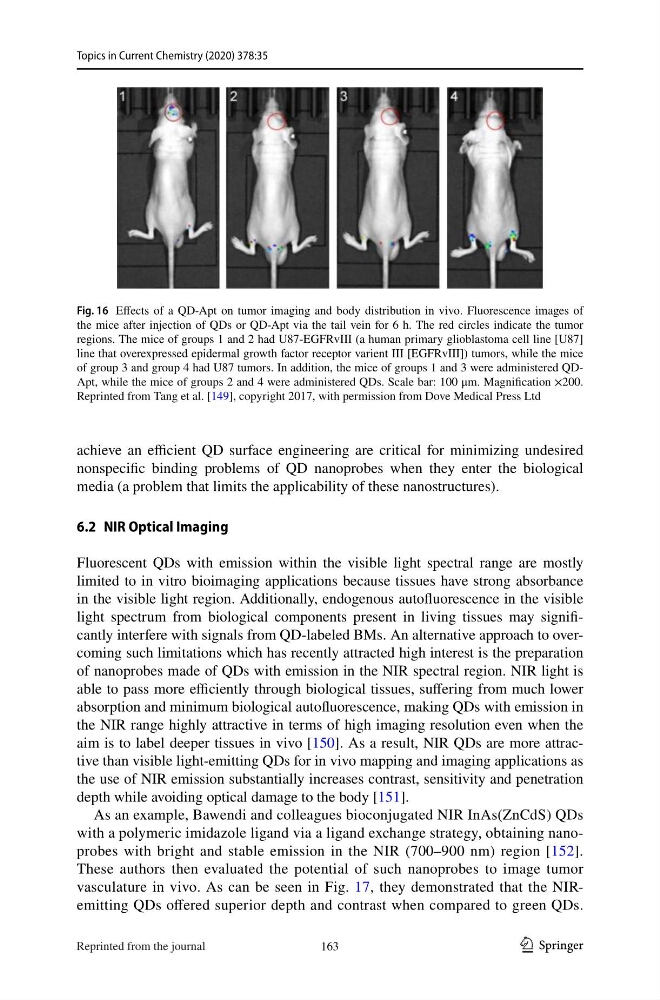
Fig. 16 Effects of a QD-Apt on tumor imaging and body distribution in vivo. Fluorescence images of
the mice after injection of QDs or QD-Apt via the tail vein for 6 h. The red circles indicate the tumor
regions. The mice of groups 1 and 2 had U87-EGFRvIII (a human primary glioblastoma cell line [U87]
line that overexpressed epidermal growth factor receptor varient III [EGFRvIII]) tumors, while the mice
of group 3 and group 4 had U87 tumors. In addition, the mice of groups 1 and 3 were administered QDApt, while the mice of groups 2 and 4 were administered QDs. Scale bar: 100 μm. Magnification ×200.
Reprinted from Tang et al. [149], copyright 2017, with permission from Dove Medical Press Ltd
163
Reprinted from the journal
Topics in Current Chemistry (2020) 378:35
achieve an efficient QD surface engineering are critical for minimizing undesired
nonspecific binding problems of QD nanoprobes when they enter the biological
media (a problem that limits the applicability of these nanostructures).
6.2 NIR Optical Imaging
Fluorescent QDs with emission within the visible light spectral range are mostly
limited to in vitro bioimaging applications because tissues have strong absorbance
in the visible light region. Additionally, endogenous autofluorescence in the visible
light spectrum from biological components present in living tissues may significantly interfere with signals from QD-labeled BMs. An alternative approach to overcoming such limitations which has recently attracted high interest is the preparation
of nanoprobes made of QDs with emission in the NIR spectral region. NIR light is
able to pass more efficiently through biological tissues, suffering from much lower
absorption and minimum biological autofluorescence, making QDs with emission in
the NIR range highly attractive in terms of high imaging resolution even when the
aim is to label deeper tissues in vivo [150]. As a result, NIR QDs are more attractive than visible light-emitting QDs for in vivo mapping and imaging applications as
the use of NIR emission substantially increases contrast, sensitivity and penetration
depth while avoiding optical damage to the body [151].
As an example, Bawendi and colleagues bioconjugated NIR InAs(ZnCdS) QDs
with a polymeric imidazole ligand via a ligand exchange strategy, obtaining nanoprobes with bright and stable emission in the NIR (700–900 nm) region [152].
These authors then evaluated the potential of such nanoprobes to image tumor
vasculature in vivo. As can be seen in Fig. 17, they demonstrated that the NIRemitting QDs offered superior depth and contrast when compared to green QDs.
Fig. 16 Effects of a QD-Apt on tumor imaging and body distribution in vivo. Fluorescence images of
the mice after injection of QDs or QD-Apt via the tail vein for 6 h. The red circles indicate the tumor
regions. The mice of groups 1 and 2 had U87-EGFRvIII (a human primary glioblastoma cell line [U87]
line that overexpressed epidermal growth factor receptor varient III [EGFRvIII]) tumors, while the mice
of group 3 and group 4 had U87 tumors. In addition, the mice of groups 1 and 3 were administered QDApt, while the mice of groups 2 and 4 were administered QDs. Scale bar: 100 μm. Magnification ×200.
Reprinted from Tang et al. [149], copyright 2017, with permission from Dove Medical Press Ltd
163
Reprinted from the journal