

Topics in Current Chemistry (2020) 378:28
1 3
to that of pure TiO 2 . Cholera toxin subunit B, which was used as a test analyte, was
sensitively detected in real time using the proposed PEC strategy. Ju’s group [28]
prepared a ternary compound consisting of TiO 2 nanotubes, polyaniline (PANI), and
gold NPs for a novel PEC bioassay based on LSPR. The LSPR band of the Au NPs
improved the system’s capacity to absorb light at ~ 540 nm and enhanced its photocurrent transfer efficiency. The electrochromism of PANI enhanced the ability of
the system to harvest visible light and to separate the charge. The biocompatibility of the ternary composite permitted the immobilization of lactate dehydrogenase
(LDH) and NAD
+
on the electrode, facilitating l-lactate PEC sensing with a detection limit of 0.15 μM. Hao et al. [29] synthesized Ag/TiO 2 -decorated, 3D-nitrogendoped graphene hydrogel (3DNGH) to promote PEC performance. The 3DNGH had
a porous structure and a large surface area that could accommodate a considerable
amount of Ag and TiO 2 NPs. Under light irradiation, electrons on the Ag surface
were transferred to TiO 2 . This Ag/TiO 2 /3DNGH nanocomposite exhibited a photocurrent that was approximately 60 times greater than that afforded by pristine TiO 2
NPs, and a sensitive label-free PEC thrombin aptasensor was constructed from this
nanocomposite.
The rational design of metal- and nonmetal-doped TiO 2 can reduce its band gap
and improve its response to visible light. Tang et al. [18] reported a PEC strategy
for aflatoxin B1 (AFB1) detection based on Ce-doped TiO 2 nanocube@MoSe 2
nanosheets. Ce was doped into the TiO 2 nanocubes using a one-step hydrothermal
method. The CB of the Ce-doped TiO 2 is at a lower energy than that of of TiO 2 , so
it has a smaller energy gap. MoSe 2 nanosheets were then grown on the TiO 2 nanocubes, yielding a system with a large surface area, and the resulting heterojunction
composite presented enhanced visible light absorption. Finally, the AuNPs were
introduced as a quenching label that markedly decreased the photocurrent, yielding an ultrasensitive assay for AFB1. Wei’s group [30] developed a Cu-doped
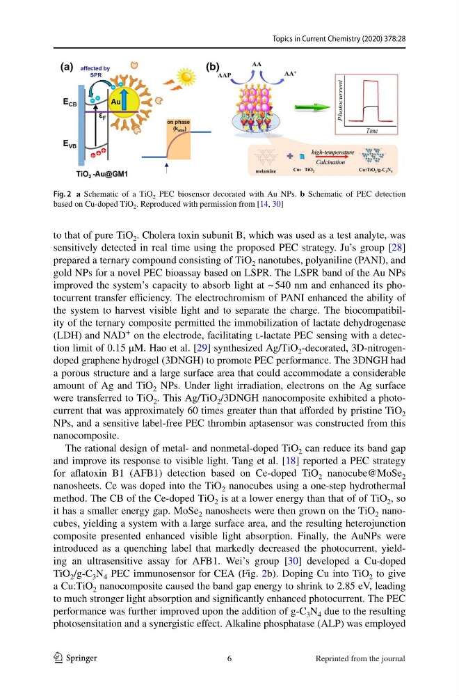
TiO 2 /g-C 3 N 4 PEC immunosensor for CEA (Fig. 2b). Doping Cu into TiO 2 to give
a Cu:TiO 2 nanocomposite caused the band gap energy to shrink to 2.85 eV, leading
to much stronger light absorption and significantly enhanced photocurrent. The PEC
performance was further improved upon the addition of g-C 3 N 4 due to the resulting
photosensitation and a synergistic effect. Alkaline phosphatase (ALP) was employed
Fig. 2 a Schematic of a TiO 2 PEC biosensor decorated with Au NPs. b Schematic of PEC detection
based on Cu-doped TiO 2 . Reproduced with permission from [14, 30]
6
Reprinted from the journal
1 3
to that of pure TiO 2 . Cholera toxin subunit B, which was used as a test analyte, was
sensitively detected in real time using the proposed PEC strategy. Ju’s group [28]
prepared a ternary compound consisting of TiO 2 nanotubes, polyaniline (PANI), and
gold NPs for a novel PEC bioassay based on LSPR. The LSPR band of the Au NPs
improved the system’s capacity to absorb light at ~ 540 nm and enhanced its photocurrent transfer efficiency. The electrochromism of PANI enhanced the ability of
the system to harvest visible light and to separate the charge. The biocompatibility of the ternary composite permitted the immobilization of lactate dehydrogenase
(LDH) and NAD
+
on the electrode, facilitating l-lactate PEC sensing with a detection limit of 0.15 μM. Hao et al. [29] synthesized Ag/TiO 2 -decorated, 3D-nitrogendoped graphene hydrogel (3DNGH) to promote PEC performance. The 3DNGH had
a porous structure and a large surface area that could accommodate a considerable
amount of Ag and TiO 2 NPs. Under light irradiation, electrons on the Ag surface
were transferred to TiO 2 . This Ag/TiO 2 /3DNGH nanocomposite exhibited a photocurrent that was approximately 60 times greater than that afforded by pristine TiO 2
NPs, and a sensitive label-free PEC thrombin aptasensor was constructed from this
nanocomposite.
The rational design of metal- and nonmetal-doped TiO 2 can reduce its band gap
and improve its response to visible light. Tang et al. [18] reported a PEC strategy
for aflatoxin B1 (AFB1) detection based on Ce-doped TiO 2 nanocube@MoSe 2
nanosheets. Ce was doped into the TiO 2 nanocubes using a one-step hydrothermal
method. The CB of the Ce-doped TiO 2 is at a lower energy than that of of TiO 2 , so
it has a smaller energy gap. MoSe 2 nanosheets were then grown on the TiO 2 nanocubes, yielding a system with a large surface area, and the resulting heterojunction
composite presented enhanced visible light absorption. Finally, the AuNPs were
introduced as a quenching label that markedly decreased the photocurrent, yielding an ultrasensitive assay for AFB1. Wei’s group [30] developed a Cu-doped
TiO 2 /g-C 3 N 4 PEC immunosensor for CEA (Fig. 2b). Doping Cu into TiO 2 to give
a Cu:TiO 2 nanocomposite caused the band gap energy to shrink to 2.85 eV, leading
to much stronger light absorption and significantly enhanced photocurrent. The PEC
performance was further improved upon the addition of g-C 3 N 4 due to the resulting
photosensitation and a synergistic effect. Alkaline phosphatase (ALP) was employed
Fig. 2 a Schematic of a TiO 2 PEC biosensor decorated with Au NPs. b Schematic of PEC detection
based on Cu-doped TiO 2 . Reproduced with permission from [14, 30]
6
Reprinted from the journal