

72
3 Mechanical Aspects of Biosystems
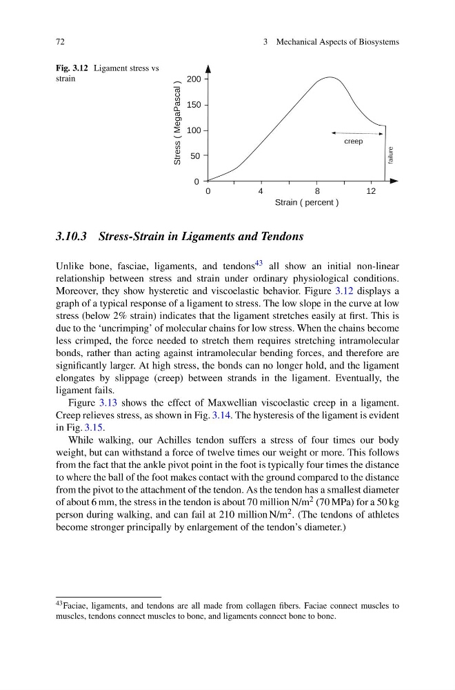
Fig. 3.12 Ligament stress vs
strain
3.10.3 Stress-Strain in Ligaments and Tendons
Unlike bone, fasciae, ligaments, and tendons 43 all show an initial non-linear
relationship between stress and strain under ordinary physiological conditions.
Moreover, they show hysteretic and viscoelastic behavior. Figure 3.12 displays a
graph of a typical response of a ligament to stress. The low slope in the curve at low
stress (below 2% strain) indicates that the ligament stretches easily at first. This is
due to the ‘uncrimping’ of molecular chains for low stress. When the chains become
less crimped, the force needed to stretch them requires stretching intramolecular
bonds, rather than acting against intramolecular bending forces, and therefore are
significantly larger. At high stress, the bonds can no longer hold, and the ligament
elongates by slippage (creep) between strands in the ligament. Eventually, the
ligament fails.
Figure 3.13 shows the effect of Maxwellian viscoelastic creep in a ligament.
Creep relieves stress, as shown in Fig. 3.14. The hysteresis of the ligament is evident
in Fig. 3.15.
While walking, our Achilles tendon suffers a stress of four times our body
weight, but can withstand a force of twelve times our weight or more. This follows
from the fact that the ankle pivot point in the foot is typically four times the distance
to where the ball of the foot makes contact with the ground compared to the distance
from the pivot to the attachment of the tendon. As the tendon has a smallest diameter
of about 6 mm, the stress in the tendon is about 70 million N/m 2 (70 MPa) for a 50 kg
person during walking, and can fail at 210 million N/m 2 . (The tendons of athletes
become stronger principally by enlargement of the tendon’s diameter.)
43 Faciae, ligaments, and tendons are all made from collagen fibers. Faciae connect muscles to
muscles, tendons connect muscles to bone, and ligaments connect bone to bone.
3 Mechanical Aspects of Biosystems
Fig. 3.12 Ligament stress vs
strain
3.10.3 Stress-Strain in Ligaments and Tendons
Unlike bone, fasciae, ligaments, and tendons 43 all show an initial non-linear
relationship between stress and strain under ordinary physiological conditions.
Moreover, they show hysteretic and viscoelastic behavior. Figure 3.12 displays a
graph of a typical response of a ligament to stress. The low slope in the curve at low
stress (below 2% strain) indicates that the ligament stretches easily at first. This is
due to the ‘uncrimping’ of molecular chains for low stress. When the chains become
less crimped, the force needed to stretch them requires stretching intramolecular
bonds, rather than acting against intramolecular bending forces, and therefore are
significantly larger. At high stress, the bonds can no longer hold, and the ligament
elongates by slippage (creep) between strands in the ligament. Eventually, the
ligament fails.
Figure 3.13 shows the effect of Maxwellian viscoelastic creep in a ligament.
Creep relieves stress, as shown in Fig. 3.14. The hysteresis of the ligament is evident
in Fig. 3.15.
While walking, our Achilles tendon suffers a stress of four times our body
weight, but can withstand a force of twelve times our weight or more. This follows
from the fact that the ankle pivot point in the foot is typically four times the distance
to where the ball of the foot makes contact with the ground compared to the distance
from the pivot to the attachment of the tendon. As the tendon has a smallest diameter
of about 6 mm, the stress in the tendon is about 70 million N/m 2 (70 MPa) for a 50 kg
person during walking, and can fail at 210 million N/m 2 . (The tendons of athletes
become stronger principally by enlargement of the tendon’s diameter.)
43 Faciae, ligaments, and tendons are all made from collagen fibers. Faciae connect muscles to
muscles, tendons connect muscles to bone, and ligaments connect bone to bone.