

10.8 The Partition Function for Ensembles
481
Tertiary Protein Structure
We now face insurmountable opportunities.
— Walt Kelly, cartoonist
In cells, proteins are constructed by ribosomes that attach amino acids together
into a sequence according to a template originating from a code on our DNA. As the
peptide chain is created, it begins to form into a secondary configuration and then
into its biological tertiary structure. In some cases, the terminal or side chains of the
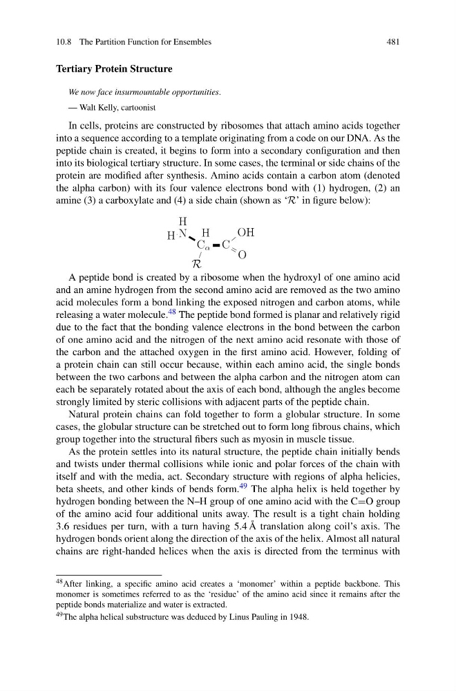
protein are modified after synthesis. Amino acids contain a carbon atom (denoted
the alpha carbon) with its four valence electrons bond with (1) hydrogen, (2) an
amine (3) a carboxylate and (4) a side chain (shown as ‘R’ in figure below):
A peptide bond is created by a ribosome when the hydroxyl of one amino acid
and an amine hydrogen from the second amino acid are removed as the two amino
acid molecules form a bond linking the exposed nitrogen and carbon atoms, while
releasing a water molecule. 48 The peptide bond formed is planar and relatively rigid
due to the fact that the bonding valence electrons in the bond between the carbon
of one amino acid and the nitrogen of the next amino acid resonate with those of
the carbon and the attached oxygen in the first amino acid. However, folding of
a protein chain can still occur because, within each amino acid, the single bonds
between the two carbons and between the alpha carbon and the nitrogen atom can
each be separately rotated about the axis of each bond, although the angles become
strongly limited by steric collisions with adjacent parts of the peptide chain.
Natural protein chains can fold together to form a globular structure. In some
cases, the globular structure can be stretched out to form long fibrous chains, which
group together into the structural fibers such as myosin in muscle tissue.
As the protein settles into its natural structure, the peptide chain initially bends
and twists under thermal collisions while ionic and polar forces of the chain with
itself and with the media, act. Secondary structure with regions of alpha helicies,
beta sheets, and other kinds of bends form. 49 The alpha helix is held together by
hydrogen bonding between the N–H group of one amino acid with the C=O group
of the amino acid four additional units away. The result is a tight chain holding
3.6 residues per turn, with a turn having 5.4 Å translation along coil’s axis. The
hydrogen bonds orient along the direction of the axis of the helix. Almost all natural
chains are right-handed helices when the axis is directed from the terminus with
48 After linking, a specific amino acid creates a ‘monomer’ within a peptide backbone. This
monomer is sometimes referred to as the ‘residue’ of the amino acid since it remains after the
peptide bonds materialize and water is extracted.
49 The alpha helical substructure was deduced by Linus Pauling in 1948.
481
Tertiary Protein Structure
We now face insurmountable opportunities.
— Walt Kelly, cartoonist
In cells, proteins are constructed by ribosomes that attach amino acids together
into a sequence according to a template originating from a code on our DNA. As the
peptide chain is created, it begins to form into a secondary configuration and then
into its biological tertiary structure. In some cases, the terminal or side chains of the
protein are modified after synthesis. Amino acids contain a carbon atom (denoted
the alpha carbon) with its four valence electrons bond with (1) hydrogen, (2) an
amine (3) a carboxylate and (4) a side chain (shown as ‘R’ in figure below):
A peptide bond is created by a ribosome when the hydroxyl of one amino acid
and an amine hydrogen from the second amino acid are removed as the two amino
acid molecules form a bond linking the exposed nitrogen and carbon atoms, while
releasing a water molecule. 48 The peptide bond formed is planar and relatively rigid
due to the fact that the bonding valence electrons in the bond between the carbon
of one amino acid and the nitrogen of the next amino acid resonate with those of
the carbon and the attached oxygen in the first amino acid. However, folding of
a protein chain can still occur because, within each amino acid, the single bonds
between the two carbons and between the alpha carbon and the nitrogen atom can
each be separately rotated about the axis of each bond, although the angles become
strongly limited by steric collisions with adjacent parts of the peptide chain.
Natural protein chains can fold together to form a globular structure. In some
cases, the globular structure can be stretched out to form long fibrous chains, which
group together into the structural fibers such as myosin in muscle tissue.
As the protein settles into its natural structure, the peptide chain initially bends
and twists under thermal collisions while ionic and polar forces of the chain with
itself and with the media, act. Secondary structure with regions of alpha helicies,
beta sheets, and other kinds of bends form. 49 The alpha helix is held together by
hydrogen bonding between the N–H group of one amino acid with the C=O group
of the amino acid four additional units away. The result is a tight chain holding
3.6 residues per turn, with a turn having 5.4 Å translation along coil’s axis. The
hydrogen bonds orient along the direction of the axis of the helix. Almost all natural
chains are right-handed helices when the axis is directed from the terminus with
48 After linking, a specific amino acid creates a ‘monomer’ within a peptide backbone. This
monomer is sometimes referred to as the ‘residue’ of the amino acid since it remains after the
peptide bonds materialize and water is extracted.
49 The alpha helical substructure was deduced by Linus Pauling in 1948.