

8.8 The Electron Microscope
305
8.8 The Electron Microscope
An electron microscope is a device capable of forming images at the nanoscale
using the fact that electron waves can have a much shorter wavelength than visible
light, and therefore allowing the microscope to have a much higher resolution, and
the fact that electron beams in a vacuum can be focused with electric and magnetic
fields (Fig. 8.13). The wavelength of an electron wave is given by the DeBroglie
relation λ = h/p, where h is Planck’s constant (6.626 × 10 −34 Joule-s) and p = mv
is the electron’s momentum. In both the optical and the electron-beam case, the
resolution of an image formed by single-wavelength waves was given by Rayleigh
as the smallest angle between two points on the object which can be resolved: R ≈
1.22 λ/d, with d the diameter of the focusing device. If f is the focal length of
the focusing device, then the smallest size one can see will be of length ≈
1.22(f/d) λ. (There is further discussion of the Rayleigh limit in Sect. 7.5.)
There are disadvantages. First, specimens should be dead, dried, and perhaps
dressed with a gold coat, 10 before being placed in a vacuum. Second, electrons
do not penetrate far in materials unless they carry energies which will break up
molecules. Third, the electron microscope tends to be big and expensive in order to
hold all the devices needed to make and sustain a good vacuum, and to generate,
focus, and detect streams of electrons in that vacuum (Fig. 8.13).
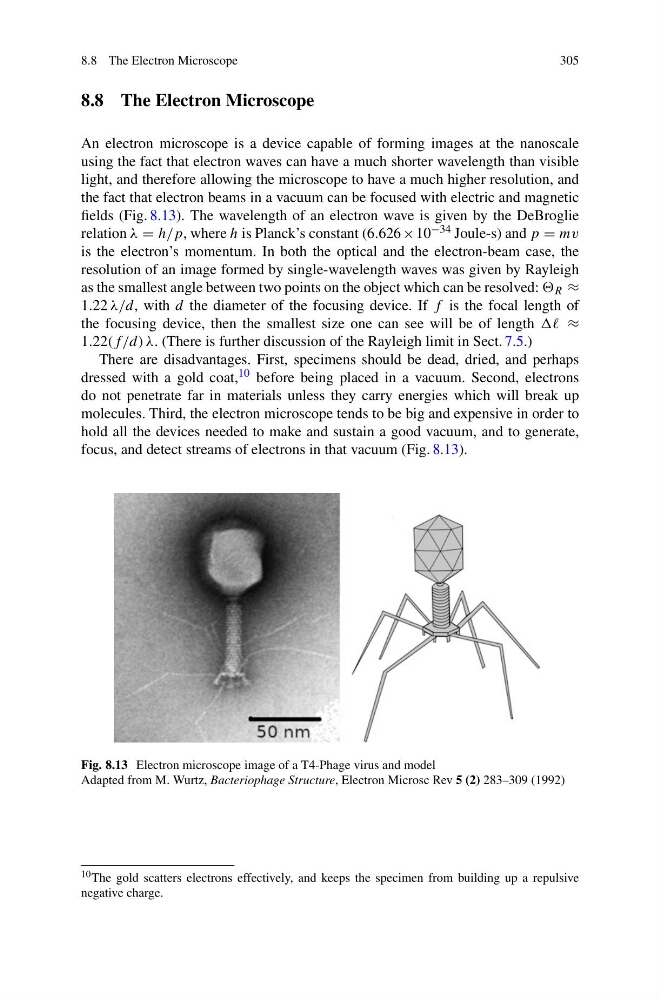
Fig. 8.13 Electron microscope image of a T4-Phage virus and model
Adapted from M. Wurtz, Bacteriophage Structure, Electron Microsc Rev 5 (2) 283–309 (1992)
10 The gold scatters electrons effectively, and keeps the specimen from building up a repulsive
negative charge.
305
8.8 The Electron Microscope
An electron microscope is a device capable of forming images at the nanoscale
using the fact that electron waves can have a much shorter wavelength than visible
light, and therefore allowing the microscope to have a much higher resolution, and
the fact that electron beams in a vacuum can be focused with electric and magnetic
fields (Fig. 8.13). The wavelength of an electron wave is given by the DeBroglie
relation λ = h/p, where h is Planck’s constant (6.626 × 10 −34 Joule-s) and p = mv
is the electron’s momentum. In both the optical and the electron-beam case, the
resolution of an image formed by single-wavelength waves was given by Rayleigh
as the smallest angle between two points on the object which can be resolved: R ≈
1.22 λ/d, with d the diameter of the focusing device. If f is the focal length of
the focusing device, then the smallest size one can see will be of length ≈
1.22(f/d) λ. (There is further discussion of the Rayleigh limit in Sect. 7.5.)
There are disadvantages. First, specimens should be dead, dried, and perhaps
dressed with a gold coat, 10 before being placed in a vacuum. Second, electrons
do not penetrate far in materials unless they carry energies which will break up
molecules. Third, the electron microscope tends to be big and expensive in order to
hold all the devices needed to make and sustain a good vacuum, and to generate,
focus, and detect streams of electrons in that vacuum (Fig. 8.13).
Fig. 8.13 Electron microscope image of a T4-Phage virus and model
Adapted from M. Wurtz, Bacteriophage Structure, Electron Microsc Rev 5 (2) 283–309 (1992)
10 The gold scatters electrons effectively, and keeps the specimen from building up a repulsive
negative charge.