

280
8 Ionizing Radiation and Life
0.3 eV), and accumulate on the surface of the crystal (where they are more stable
than in the bulk crystal). In the photographic process, a silver bromide emulsion is
prepared with micrometer or smaller grains of AgBr salt crystals in a gelatin that
is spread in a dark room on a surface such as glass. After exposure to radiation,
the film is placed in a ‘developer’ solution to release the metallic silver from
the exposed crystals. After development, a ‘fixer’ solution (sodium thiosulfate)
dissolves the unexposed silver bromide, and the film is then washed. Now the film
will have black silver where light has reached the film, and be clear where it has
not. On 8 Nov 1895, Wilhelm Röentgen noticed that barium platinocyanide gave
off a faint flickering light near a Crookes tube. 1 He named the suspected unknown
emissions from the Crookes tube ‘X-rays’ (‘X’ for unknown). Experimenting
with the discovery, he soon found that X-rays would penetrate human tissue and
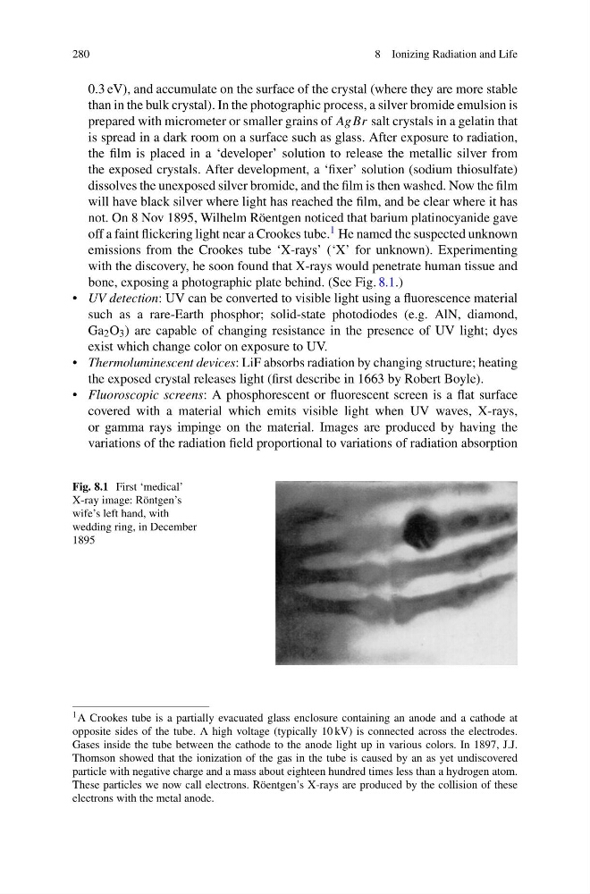
bone, exposing a photographic plate behind. (See Fig. 8.1.)
• UV detection: UV can be converted to visible light using a fluorescence material
such as a rare-Earth phosphor; solid-state photodiodes (e.g. AlN, diamond,
Ga 2 O 3 ) are capable of changing resistance in the presence of UV light; dyes
exist which change color on exposure to UV.
• Thermoluminescent devices: LiF absorbs radiation by changing structure; heating
the exposed crystal releases light (first describe in 1663 by Robert Boyle).
• Fluoroscopic screens: A phosphorescent or fluorescent screen is a flat surface
covered with a material which emits visible light when UV waves, X-rays,
or gamma rays impinge on the material. Images are produced by having the
variations of the radiation field proportional to variations of radiation absorption
Fig. 8.1 First ‘medical’
X-ray image: Röntgen’s
wife’s left hand, with
wedding ring, in December
1895
1 A Crookes tube is a partially evacuated glass enclosure containing an anode and a cathode at
opposite sides of the tube. A high voltage (typically 10 kV) is connected across the electrodes.
Gases inside the tube between the cathode to the anode light up in various colors. In 1897, J.J.
Thomson showed that the ionization of the gas in the tube is caused by an as yet undiscovered
particle with negative charge and a mass about eighteen hundred times less than a hydrogen atom.
These particles we now call electrons. Röentgen’s X-rays are produced by the collision of these
electrons with the metal anode.
8 Ionizing Radiation and Life
0.3 eV), and accumulate on the surface of the crystal (where they are more stable
than in the bulk crystal). In the photographic process, a silver bromide emulsion is
prepared with micrometer or smaller grains of AgBr salt crystals in a gelatin that
is spread in a dark room on a surface such as glass. After exposure to radiation,
the film is placed in a ‘developer’ solution to release the metallic silver from
the exposed crystals. After development, a ‘fixer’ solution (sodium thiosulfate)
dissolves the unexposed silver bromide, and the film is then washed. Now the film
will have black silver where light has reached the film, and be clear where it has
not. On 8 Nov 1895, Wilhelm Röentgen noticed that barium platinocyanide gave
off a faint flickering light near a Crookes tube. 1 He named the suspected unknown
emissions from the Crookes tube ‘X-rays’ (‘X’ for unknown). Experimenting
with the discovery, he soon found that X-rays would penetrate human tissue and
bone, exposing a photographic plate behind. (See Fig. 8.1.)
• UV detection: UV can be converted to visible light using a fluorescence material
such as a rare-Earth phosphor; solid-state photodiodes (e.g. AlN, diamond,
Ga 2 O 3 ) are capable of changing resistance in the presence of UV light; dyes
exist which change color on exposure to UV.
• Thermoluminescent devices: LiF absorbs radiation by changing structure; heating
the exposed crystal releases light (first describe in 1663 by Robert Boyle).
• Fluoroscopic screens: A phosphorescent or fluorescent screen is a flat surface
covered with a material which emits visible light when UV waves, X-rays,
or gamma rays impinge on the material. Images are produced by having the
variations of the radiation field proportional to variations of radiation absorption
Fig. 8.1 First ‘medical’
X-ray image: Röntgen’s
wife’s left hand, with
wedding ring, in December
1895
1 A Crookes tube is a partially evacuated glass enclosure containing an anode and a cathode at
opposite sides of the tube. A high voltage (typically 10 kV) is connected across the electrodes.
Gases inside the tube between the cathode to the anode light up in various colors. In 1897, J.J.
Thomson showed that the ionization of the gas in the tube is caused by an as yet undiscovered
particle with negative charge and a mass about eighteen hundred times less than a hydrogen atom.
These particles we now call electrons. Röentgen’s X-rays are produced by the collision of these
electrons with the metal anode.