

7.27 Vision and the Eye
259
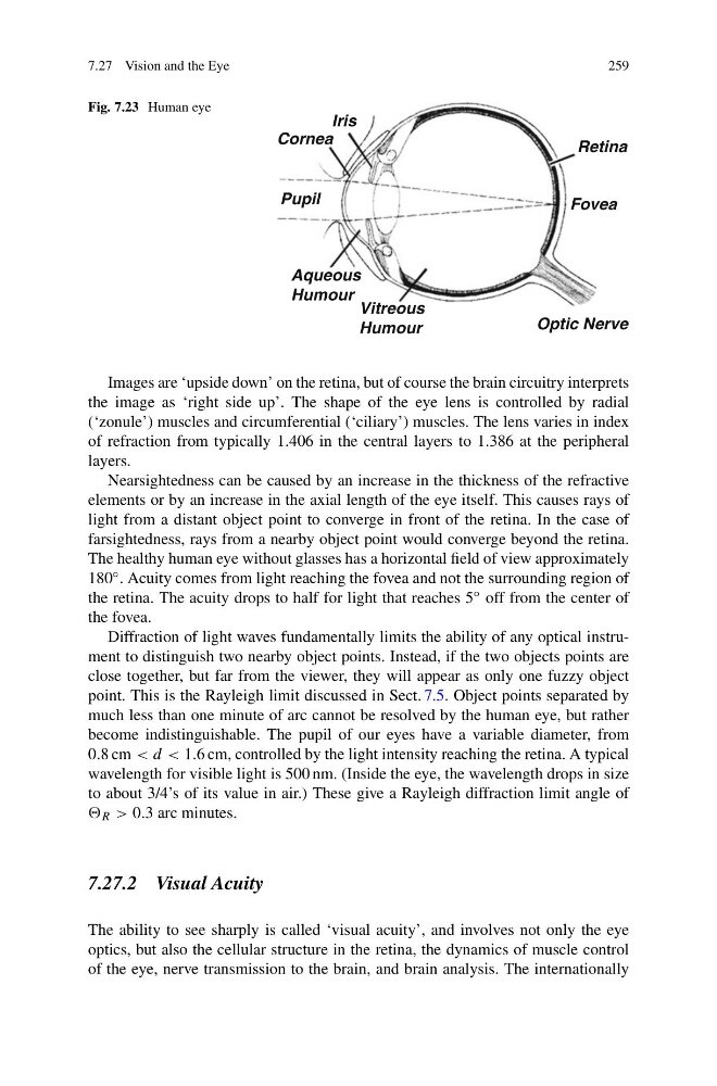
Fig. 7.23 Human eye
Retina
Fovea
Optic Nerve
Vitreous
Humour
Aqueous
Humour
Pupil
Iris
Cornea
Images are ‘upside down’ on the retina, but of course the brain circuitry interprets
the image as ‘right side up’. The shape of the eye lens is controlled by radial
(‘zonule’) muscles and circumferential (‘ciliary’) muscles. The lens varies in index
of refraction from typically 1.406 in the central layers to 1.386 at the peripheral
layers.
Nearsightedness can be caused by an increase in the thickness of the refractive
elements or by an increase in the axial length of the eye itself. This causes rays of
light from a distant object point to converge in front of the retina. In the case of
farsightedness, rays from a nearby object point would converge beyond the retina.
The healthy human eye without glasses has a horizontal field of view approximately
180 ◦ . Acuity comes from light reaching the fovea and not the surrounding region of
the retina. The acuity drops to half for light that reaches 5 ◦ off from the center of
the fovea.
Diffraction of light waves fundamentally limits the ability of any optical instrument to distinguish two nearby object points. Instead, if the two objects points are
close together, but far from the viewer, they will appear as only one fuzzy object
point. This is the Rayleigh limit discussed in Sect. 7.5. Object points separated by
much less than one minute of arc cannot be resolved by the human eye, but rather
become indistinguishable. The pupil of our eyes have a variable diameter, from
0.8 cm < d < 1.6 cm, controlled by the light intensity reaching the retina. A typical
wavelength for visible light is 500 nm. (Inside the eye, the wavelength drops in size
to about 3/4’s of its value in air.) These give a Rayleigh diffraction limit angle of
R > 0.3 arc minutes.
7.27.2 Visual Acuity
The ability to see sharply is called ‘visual acuity’, and involves not only the eye
optics, but also the cellular structure in the retina, the dynamics of muscle control
of the eye, nerve transmission to the brain, and brain analysis. The internationally
259
Fig. 7.23 Human eye
Retina
Fovea
Optic Nerve
Vitreous
Humour
Aqueous
Humour
Pupil
Iris
Cornea
Images are ‘upside down’ on the retina, but of course the brain circuitry interprets
the image as ‘right side up’. The shape of the eye lens is controlled by radial
(‘zonule’) muscles and circumferential (‘ciliary’) muscles. The lens varies in index
of refraction from typically 1.406 in the central layers to 1.386 at the peripheral
layers.
Nearsightedness can be caused by an increase in the thickness of the refractive
elements or by an increase in the axial length of the eye itself. This causes rays of
light from a distant object point to converge in front of the retina. In the case of
farsightedness, rays from a nearby object point would converge beyond the retina.
The healthy human eye without glasses has a horizontal field of view approximately
180 ◦ . Acuity comes from light reaching the fovea and not the surrounding region of
the retina. The acuity drops to half for light that reaches 5 ◦ off from the center of
the fovea.
Diffraction of light waves fundamentally limits the ability of any optical instrument to distinguish two nearby object points. Instead, if the two objects points are
close together, but far from the viewer, they will appear as only one fuzzy object
point. This is the Rayleigh limit discussed in Sect. 7.5. Object points separated by
much less than one minute of arc cannot be resolved by the human eye, but rather
become indistinguishable. The pupil of our eyes have a variable diameter, from
0.8 cm < d < 1.6 cm, controlled by the light intensity reaching the retina. A typical
wavelength for visible light is 500 nm. (Inside the eye, the wavelength drops in size
to about 3/4’s of its value in air.) These give a Rayleigh diffraction limit angle of
R > 0.3 arc minutes.
7.27.2 Visual Acuity
The ability to see sharply is called ‘visual acuity’, and involves not only the eye
optics, but also the cellular structure in the retina, the dynamics of muscle control
of the eye, nerve transmission to the brain, and brain analysis. The internationally