

232
7 Light in Biology and Medicine
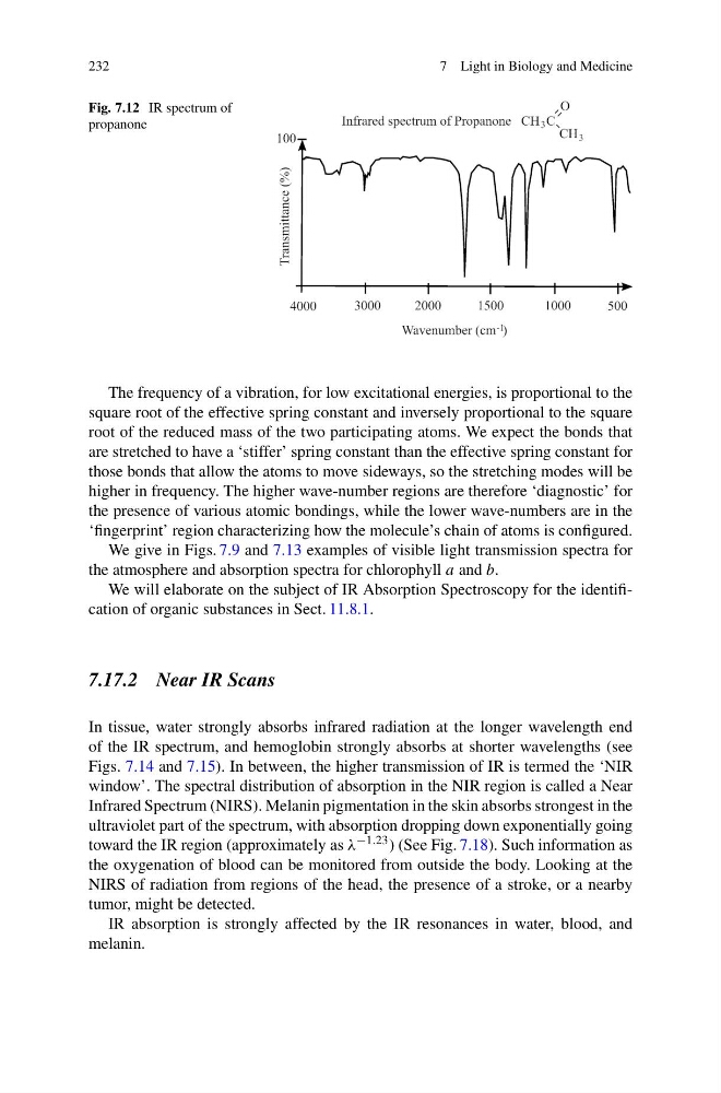
Fig. 7.12 IR spectrum of
propanone
The frequency of a vibration, for low excitational energies, is proportional to the
square root of the effective spring constant and inversely proportional to the square
root of the reduced mass of the two participating atoms. We expect the bonds that
are stretched to have a ‘stiffer’ spring constant than the effective spring constant for
those bonds that allow the atoms to move sideways, so the stretching modes will be
higher in frequency. The higher wave-number regions are therefore ‘diagnostic’ for
the presence of various atomic bondings, while the lower wave-numbers are in the
‘fingerprint’ region characterizing how the molecule’s chain of atoms is configured.
We give in Figs. 7.9 and 7.13 examples of visible light transmission spectra for
the atmosphere and absorption spectra for chlorophyll a and b.
We will elaborate on the subject of IR Absorption Spectroscopy for the identification of organic substances in Sect. 11.8.1.
7.17.2 Near IR Scans
In tissue, water strongly absorbs infrared radiation at the longer wavelength end
of the IR spectrum, and hemoglobin strongly absorbs at shorter wavelengths (see
Figs. 7.14 and 7.15). In between, the higher transmission of IR is termed the ‘NIR
window’. The spectral distribution of absorption in the NIR region is called a Near
Infrared Spectrum (NIRS). Melanin pigmentation in the skin absorbs strongest in the
ultraviolet part of the spectrum, with absorption dropping down exponentially going
toward the IR region (approximately as λ −1.23 ) (See Fig. 7.18). Such information as
the oxygenation of blood can be monitored from outside the body. Looking at the
NIRS of radiation from regions of the head, the presence of a stroke, or a nearby
tumor, might be detected.
IR absorption is strongly affected by the IR resonances in water, blood, and
melanin.
7 Light in Biology and Medicine
Fig. 7.12 IR spectrum of
propanone
The frequency of a vibration, for low excitational energies, is proportional to the
square root of the effective spring constant and inversely proportional to the square
root of the reduced mass of the two participating atoms. We expect the bonds that
are stretched to have a ‘stiffer’ spring constant than the effective spring constant for
those bonds that allow the atoms to move sideways, so the stretching modes will be
higher in frequency. The higher wave-number regions are therefore ‘diagnostic’ for
the presence of various atomic bondings, while the lower wave-numbers are in the
‘fingerprint’ region characterizing how the molecule’s chain of atoms is configured.
We give in Figs. 7.9 and 7.13 examples of visible light transmission spectra for
the atmosphere and absorption spectra for chlorophyll a and b.
We will elaborate on the subject of IR Absorption Spectroscopy for the identification of organic substances in Sect. 11.8.1.
7.17.2 Near IR Scans
In tissue, water strongly absorbs infrared radiation at the longer wavelength end
of the IR spectrum, and hemoglobin strongly absorbs at shorter wavelengths (see
Figs. 7.14 and 7.15). In between, the higher transmission of IR is termed the ‘NIR
window’. The spectral distribution of absorption in the NIR region is called a Near
Infrared Spectrum (NIRS). Melanin pigmentation in the skin absorbs strongest in the
ultraviolet part of the spectrum, with absorption dropping down exponentially going
toward the IR region (approximately as λ −1.23 ) (See Fig. 7.18). Such information as
the oxygenation of blood can be monitored from outside the body. Looking at the
NIRS of radiation from regions of the head, the presence of a stroke, or a nearby
tumor, might be detected.
IR absorption is strongly affected by the IR resonances in water, blood, and
melanin.