

scattering. Qualitatively the scattering of X-rays results from the oscillation of the
electrons in atoms which interact with the incoming electromagnetic waves of
X-rays. Atoms with higher atomic numbers scatter more effectively because they
have more electrons circulating the nucleus. The extent of scattering depends not
only on the electron density distribution but also on the angle between the incoming
X-rays and the diffraction plane, i.e. the scattering is larger in the forward direction
and smaller in the reverse direction. The distribution of electron density in the plane
involved in the Bragg reflection is expressed using a Fourier analysis which depends
on the co-ordinates of the atoms and the scattering factors of the atoms located in that
plane. These are summed over all the atoms in the unit cell, and the quantity is
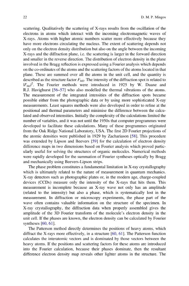
described as the structure factor F hkl . The intensity of the diffraction spot is related to
|F hkl |
2 . The Fourier methods were introduced in 1925 by W. Duane and
R.J. Havighurst [56–57] who also modelled the thermal vibrations of the atoms.
The measurement of the integrated intensities of the diffraction spots became
possible either from the photographic data or by using more sophisticated X-ray
measurements. Least squares methods were also developed in order to refine at the
positional and thermal parameters and minimise the difference between the calculated and observed intensities. Initially the complexity of the calculations limited the
number of variables, and it was not until the 1950s that computer programmes were
developed to facilitate these calculations. Many of these programmes originated
from the Oak Ridge National Laboratory, USA. The first 2D Fourier projections of
the atomic densities were published in 1929 by Zachariason [58]. This procedure
was extended by Lipson and Beevers [59] for the calculation of electron density
difference maps in two dimensions based on Fourier analysis which proved particularly useful for solving the structures of organic molecules. Numerical methods
were rapidly developed for the summation of Fourier syntheses optically by Bragg
and mechanically using Beevers-Lipson strips.
The phase problem constitutes a fundamental limitation in X-ray crystallography
which is ultimately related to the nature of measurement in quantum mechanics.
X-ray detectors such as photographic plates or, in the modern age, charge-coupled
devices (CCDs) measure only the intensity of the X-rays that hits them. This
measurement is incomplete because an X-ray wave not only has an amplitude
(related to the intensity) but also a phase, which is systematically lost in the
measurement. In diffraction or microscopy experiments, the phase part of the
wave often contains valuable information on the structure of the specimen. In
X-ray crystallography, the diffraction data when properly assembled gives the
amplitude of the 3D Fourier transform of the molecule’s electron density in the
unit cell. If the phases are known, the electron density can be calculated by Fourier
syntheses [60, 61].
The Patterson method directly determines the positions of heavy atoms, which
diffract the X-rays more effectively, in a structure [60, 61]. The Patterson function
calculates the interatomic vectors and is dominated by those vectors between the
heavy atoms. If the positions and scattering factors for these atoms are introduced
into the Fourier calculation, because their phases dominate, then the resultant
difference electron density map reveals other lighter atoms in the structure. The
22
D. M. P. Mingos
electrons in atoms which interact with the incoming electromagnetic waves of
X-rays. Atoms with higher atomic numbers scatter more effectively because they
have more electrons circulating the nucleus. The extent of scattering depends not
only on the electron density distribution but also on the angle between the incoming
X-rays and the diffraction plane, i.e. the scattering is larger in the forward direction
and smaller in the reverse direction. The distribution of electron density in the plane
involved in the Bragg reflection is expressed using a Fourier analysis which depends
on the co-ordinates of the atoms and the scattering factors of the atoms located in that
plane. These are summed over all the atoms in the unit cell, and the quantity is
described as the structure factor F hkl . The intensity of the diffraction spot is related to
|F hkl |
2 . The Fourier methods were introduced in 1925 by W. Duane and
R.J. Havighurst [56–57] who also modelled the thermal vibrations of the atoms.
The measurement of the integrated intensities of the diffraction spots became
possible either from the photographic data or by using more sophisticated X-ray
measurements. Least squares methods were also developed in order to refine at the
positional and thermal parameters and minimise the difference between the calculated and observed intensities. Initially the complexity of the calculations limited the
number of variables, and it was not until the 1950s that computer programmes were
developed to facilitate these calculations. Many of these programmes originated
from the Oak Ridge National Laboratory, USA. The first 2D Fourier projections of
the atomic densities were published in 1929 by Zachariason [58]. This procedure
was extended by Lipson and Beevers [59] for the calculation of electron density
difference maps in two dimensions based on Fourier analysis which proved particularly useful for solving the structures of organic molecules. Numerical methods
were rapidly developed for the summation of Fourier syntheses optically by Bragg
and mechanically using Beevers-Lipson strips.
The phase problem constitutes a fundamental limitation in X-ray crystallography
which is ultimately related to the nature of measurement in quantum mechanics.
X-ray detectors such as photographic plates or, in the modern age, charge-coupled
devices (CCDs) measure only the intensity of the X-rays that hits them. This
measurement is incomplete because an X-ray wave not only has an amplitude
(related to the intensity) but also a phase, which is systematically lost in the
measurement. In diffraction or microscopy experiments, the phase part of the
wave often contains valuable information on the structure of the specimen. In
X-ray crystallography, the diffraction data when properly assembled gives the
amplitude of the 3D Fourier transform of the molecule’s electron density in the
unit cell. If the phases are known, the electron density can be calculated by Fourier
syntheses [60, 61].
The Patterson method directly determines the positions of heavy atoms, which
diffract the X-rays more effectively, in a structure [60, 61]. The Patterson function
calculates the interatomic vectors and is dominated by those vectors between the
heavy atoms. If the positions and scattering factors for these atoms are introduced
into the Fourier calculation, because their phases dominate, then the resultant
difference electron density map reveals other lighter atoms in the structure. The
22
D. M. P. Mingos