

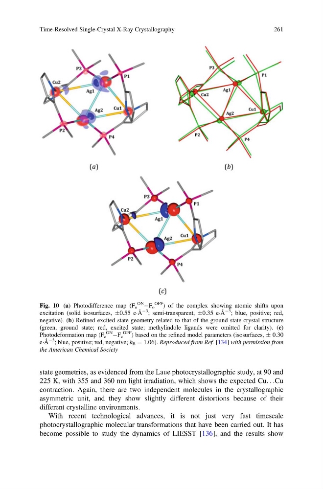
state geometries, as evidenced from the Laue photocrystallographic study, at 90 and
225 K, with 355 and 360 nm light irradiation, which shows the expected Cu. . .Cu
contraction. Again, there are two independent molecules in the crystallographic
asymmetric unit, and they show slightly different distortions because of their
different crystalline environments.
With recent technological advances, it is not just very fast timescale
photocrystallographic molecular transformations that have been carried out. It has
become possible to study the dynamics of LIESST [136], and the results show
Fig. 10 (a) Photodifference map (F o
ON ÀF o
OFF
) of the complex showing atomic shifts upon
excitation (solid isosurfaces, Æ0.55 eÁÅ
À3
; semi-transparent, Æ0.35 eÁÅ
À3
; blue, positive; red,
negative). (b) Refined excited state geometry related to that of the ground state crystal structure
(green, ground state; red, excited state; methylindole ligands were omitted for clarity). (c)
Photodeformation map (F c
ON ÀF c
OFF
) based on the refined model parameters (isosurfaces, Æ 0.30
eÁÅ
À3
; blue, positive; red, negative; k B ¼ 1.06). Reproduced from Ref. [134] with permission from
the American Chemical Society
Time-Resolved Single-Crystal X-Ray Crystallography
261
225 K, with 355 and 360 nm light irradiation, which shows the expected Cu. . .Cu
contraction. Again, there are two independent molecules in the crystallographic
asymmetric unit, and they show slightly different distortions because of their
different crystalline environments.
With recent technological advances, it is not just very fast timescale
photocrystallographic molecular transformations that have been carried out. It has
become possible to study the dynamics of LIESST [136], and the results show
Fig. 10 (a) Photodifference map (F o
ON ÀF o
OFF
) of the complex showing atomic shifts upon
excitation (solid isosurfaces, Æ0.55 eÁÅ
À3
; semi-transparent, Æ0.35 eÁÅ
À3
; blue, positive; red,
negative). (b) Refined excited state geometry related to that of the ground state crystal structure
(green, ground state; red, excited state; methylindole ligands were omitted for clarity). (c)
Photodeformation map (F c
ON ÀF c
OFF
) based on the refined model parameters (isosurfaces, Æ 0.30
eÁÅ
À3
; blue, positive; red, negative; k B ¼ 1.06). Reproduced from Ref. [134] with permission from
the American Chemical Society
Time-Resolved Single-Crystal X-Ray Crystallography
261