

and a focal spot of 4 μm is irradiated on the foam at the intensity 3.5 Â 10
20 W/cm
2
(a 0 ¼ 18). The target is of 8 μm thick porous foam on 0.8 μm thick aluminum foils
(n ¼ 40n c ) with 0.02 μm low-density (10n c ) CH layer on the rear surface.
The laser is irradiated with p-polarization as shown. It is noted that the space
dimension is about 50 Â 20 μm [19]. In Fig. 7.28b, the electron density distribution
is shown at time 67 fs during the laser heating. It is seen that a relatively wide range
about 20 μm diameter is ionized, although the laser-focusing spot is 4 μm. The
irradiated laser is scattered and bended by the porous structure and seems to diffuse
to the surrounding region. Figure 7.28b is at t ¼ 134 fs, after the laser irradiation, and
the density structure of the foam layer becomes almost uniform. It is concluded in
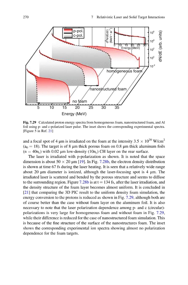
[21] that comparing the 3D PIC result to the uniform density foam simulation, the
energy conversion to the protons is reduced as shown in Fig. 7.29, although both are
of course better than the case without foam layer on the aluminum foil. It is also
necessary to note that the laser polarization dependence among p- and c (circular)polarizations is very large for homogeneous foam and without foam in Fig. 7.29,
while their difference is reduced for the case of nanostructured foam simulation. This
is because of the fine structure of the surface of the nanostructures foam. The inset
shows the corresponding experimental ion spectra showing almost no polarization
dependence for the foam targets.
5
10
15
20
Energy (MeV)
25
30
35
no foam
nanostructured foam
homogeneous foam
Energy (MeV)
5
d
2
N/dEdΩ
(MeV -1
sr
-1
)
10 15 20 25 30
dN/dE (arb. units)
p-pol.
c-pol.
10
-2
10
0
10
2
10
4
10
8
Fig. 7.29 Calculated proton energy spectra from homogeneous foam, nanostructured foam, and Al
foil using p- and c-polarized laser pulse. The inset shows the corresponding experimental spectra.
[Figure 5 in Ref. 21]
270
7 Relativistic Laser and Solid Target Interactions
20 W/cm
2
(a 0 ¼ 18). The target is of 8 μm thick porous foam on 0.8 μm thick aluminum foils
(n ¼ 40n c ) with 0.02 μm low-density (10n c ) CH layer on the rear surface.
The laser is irradiated with p-polarization as shown. It is noted that the space
dimension is about 50 Â 20 μm [19]. In Fig. 7.28b, the electron density distribution
is shown at time 67 fs during the laser heating. It is seen that a relatively wide range
about 20 μm diameter is ionized, although the laser-focusing spot is 4 μm. The
irradiated laser is scattered and bended by the porous structure and seems to diffuse
to the surrounding region. Figure 7.28b is at t ¼ 134 fs, after the laser irradiation, and
the density structure of the foam layer becomes almost uniform. It is concluded in
[21] that comparing the 3D PIC result to the uniform density foam simulation, the
energy conversion to the protons is reduced as shown in Fig. 7.29, although both are
of course better than the case without foam layer on the aluminum foil. It is also
necessary to note that the laser polarization dependence among p- and c (circular)polarizations is very large for homogeneous foam and without foam in Fig. 7.29,
while their difference is reduced for the case of nanostructured foam simulation. This
is because of the fine structure of the surface of the nanostructures foam. The inset
shows the corresponding experimental ion spectra showing almost no polarization
dependence for the foam targets.
5
10
15
20
Energy (MeV)
25
30
35
no foam
nanostructured foam
homogeneous foam
Energy (MeV)
5
d
2
N/dEdΩ
(MeV -1
sr
-1
)
10 15 20 25 30
dN/dE (arb. units)
p-pol.
c-pol.
10
-2
10
0
10
2
10
4
10
8
Fig. 7.29 Calculated proton energy spectra from homogeneous foam, nanostructured foam, and Al
foil using p- and c-polarized laser pulse. The inset shows the corresponding experimental spectra.
[Figure 5 in Ref. 21]
270
7 Relativistic Laser and Solid Target Interactions