

3.3 Single Crystal X-ray Diffraction (XRD) Analyses
87
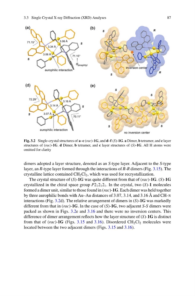
Fig. 3.2 Single-crystal structures of a–c (rac)-1G, and d–f (S)-1G. a Dimer, b tetramer, and c layer
structures of (rac)-1G. d Dimer, b tetramer, and c layer structures of (S)-1G. All H atoms were
omitted for clarity
dimers adopted a layer structure, denoted as an S-type layer. Adjacent to the S-type
layer, an R-type layer formed through the interactions of R-R dimers (Fig. 3.15). The
crystalline lattice contained CH 2 Cl 2 , which was used for recrystallization.
The crystal structure of (S)-1G was quite different from that of (rac)-1G. (S)-1G
crystallized in the chiral space group P2 1 2 1 2 1 . In the crystal, two (S)-1 molecules
formed a dimer unit, similar to those found in (rac)-1G. Each dimer was held together
by three aurophilic bonds with Au–Au distances of 3.07, 3.14, and 3.16 Å and CH-π
interactions (Fig. 3.2d). The relative arrangement of dimers in (S)-1G was markedly
different from that in (rac)-1G. In the case of (S)-1G, two adjacent S-S dimers were
packed as shown in Figs. 3.2e and 3.16 and there were no inversion centers. This
difference of dimer arrangement reflects how the layer structure of (S)-1G is distinct
from that of (rac)-1G (Figs. 3.15 and 3.16). Disordered CH 2 Cl 2 molecules were
located between the two adjacent dimers (Figs. 3.15 and 3.16).
87
Fig. 3.2 Single-crystal structures of a–c (rac)-1G, and d–f (S)-1G. a Dimer, b tetramer, and c layer
structures of (rac)-1G. d Dimer, b tetramer, and c layer structures of (S)-1G. All H atoms were
omitted for clarity
dimers adopted a layer structure, denoted as an S-type layer. Adjacent to the S-type
layer, an R-type layer formed through the interactions of R-R dimers (Fig. 3.15). The
crystalline lattice contained CH 2 Cl 2 , which was used for recrystallization.
The crystal structure of (S)-1G was quite different from that of (rac)-1G. (S)-1G
crystallized in the chiral space group P2 1 2 1 2 1 . In the crystal, two (S)-1 molecules
formed a dimer unit, similar to those found in (rac)-1G. Each dimer was held together
by three aurophilic bonds with Au–Au distances of 3.07, 3.14, and 3.16 Å and CH-π
interactions (Fig. 3.2d). The relative arrangement of dimers in (S)-1G was markedly
different from that in (rac)-1G. In the case of (S)-1G, two adjacent S-S dimers were
packed as shown in Figs. 3.2e and 3.16 and there were no inversion centers. This
difference of dimer arrangement reflects how the layer structure of (S)-1G is distinct
from that of (rac)-1G (Figs. 3.15 and 3.16). Disordered CH 2 Cl 2 molecules were
located between the two adjacent dimers (Figs. 3.15 and 3.16).