

5.7 Experimental Section
151
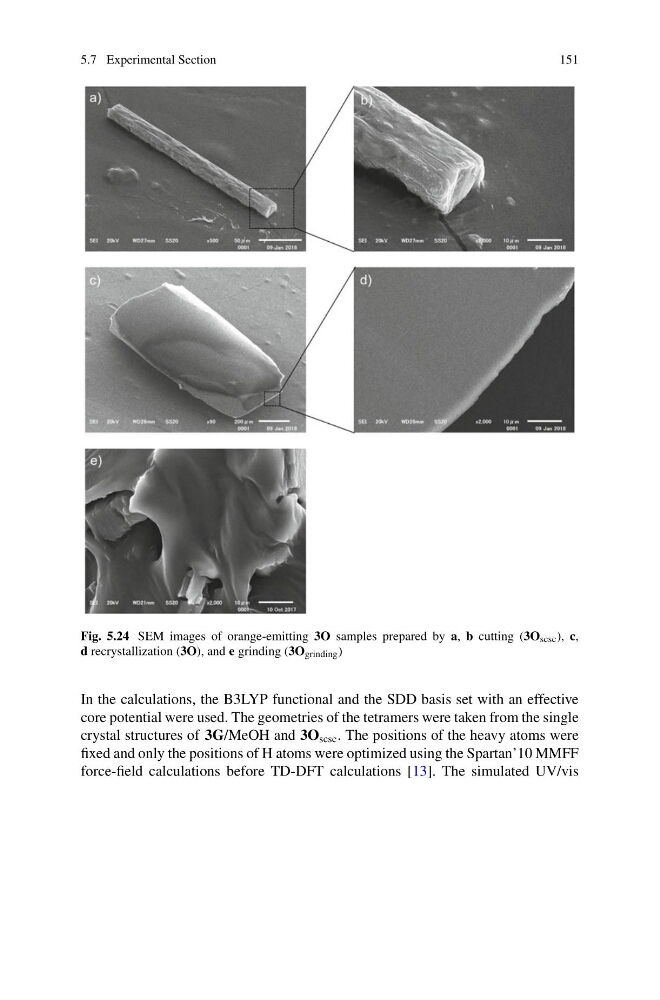
Fig. 5.24 SEM images of orange-emitting 3O samples prepared by a, b cutting (3O scsc ), c,
d recrystallization (3O), and e grinding (3O grinding )
In the calculations, the B3LYP functional and the SDD basis set with an effective
core potential were used. The geometries of the tetramers were taken from the single
crystal structures of 3G/MeOH and 3O scsc . The positions of the heavy atoms were
fixed and only the positions of H atoms were optimized using the Spartan’10 MMFF
force-field calculations before TD-DFT calculations [13]. The simulated UV/vis
151
Fig. 5.24 SEM images of orange-emitting 3O samples prepared by a, b cutting (3O scsc ), c,
d recrystallization (3O), and e grinding (3O grinding )
In the calculations, the B3LYP functional and the SDD basis set with an effective
core potential were used. The geometries of the tetramers were taken from the single
crystal structures of 3G/MeOH and 3O scsc . The positions of the heavy atoms were
fixed and only the positions of H atoms were optimized using the Spartan’10 MMFF
force-field calculations before TD-DFT calculations [13]. The simulated UV/vis