

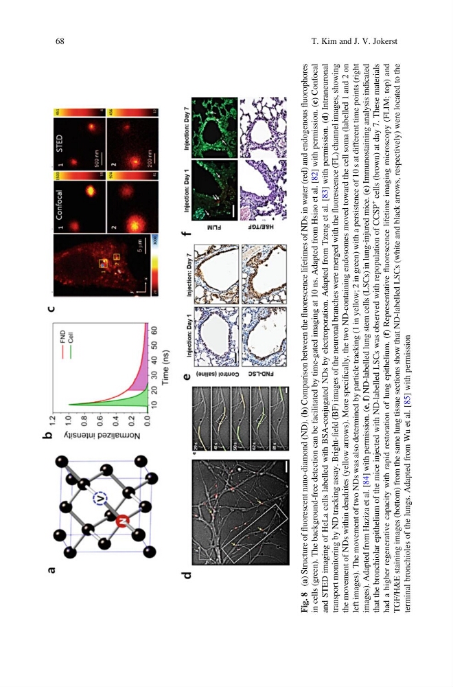
Fig. 8
(a) Structure of
fluorescent nano-diamond (ND). (b) Comparison between the
fluorescence lifetimes of NDs in water (red) and endogenous
fluorophores
in cells (green). The background-free detection can be facilitated by time-gated imaging at 10 ns. Adapted from Hsiao et al. [82] with permission. (c) Confocal
and STED imaging of HeLa cells labelled with BSA-conjugated NDs by electroporation. Adapted from Tzeng et al. [83] with permission. (d) Intraneuronal
transport monitoring by ND tracking assay. Bright-field (BF) images of the neuronal branches were merged with the
fluorescence (FL) channel images, showing
the movement of NDs within dendrites (yellow arrows). More specifically, the two ND-containing endosomes moved toward the cell soma (labelled 1 and 2 on
left images). The movement of two NDs was also determined by particle tracking (1 in yellow; 2 in green) with a persistence of 10 s at different time points (right
images). Adapted from Haziza et al. [84] with permission. (e, f)
ND-labelled lung stem cells (LSCs) in lung-injured mice. (e) Immunostaining analysis indicated
that the bronchiolar epithelium of the mice injected with ND-labelled LSCs was observed with repopulation of CCSP
+
cells (brown) at day 7. These materials
had a higher regenerative capacity with rapid restoration of lung epithelium. (f) Representative
fluorescence lifetime imaging microscopy (FLIM; top) and
TGF/H&E staining images (bottom) from the same lung tissue sections show that ND-labelled LSCs (white and black arrows, respectively) were located to the
terminal bronchioles of the lungs. Adapted from Wu et al. [85] with permission
68
T. Kim and J. V. Jokerst
(a) Structure of
fluorescent nano-diamond (ND). (b) Comparison between the
fluorescence lifetimes of NDs in water (red) and endogenous
fluorophores
in cells (green). The background-free detection can be facilitated by time-gated imaging at 10 ns. Adapted from Hsiao et al. [82] with permission. (c) Confocal
and STED imaging of HeLa cells labelled with BSA-conjugated NDs by electroporation. Adapted from Tzeng et al. [83] with permission. (d) Intraneuronal
transport monitoring by ND tracking assay. Bright-field (BF) images of the neuronal branches were merged with the
fluorescence (FL) channel images, showing
the movement of NDs within dendrites (yellow arrows). More specifically, the two ND-containing endosomes moved toward the cell soma (labelled 1 and 2 on
left images). The movement of two NDs was also determined by particle tracking (1 in yellow; 2 in green) with a persistence of 10 s at different time points (right
images). Adapted from Haziza et al. [84] with permission. (e, f)
ND-labelled lung stem cells (LSCs) in lung-injured mice. (e) Immunostaining analysis indicated
that the bronchiolar epithelium of the mice injected with ND-labelled LSCs was observed with repopulation of CCSP
+
cells (brown) at day 7. These materials
had a higher regenerative capacity with rapid restoration of lung epithelium. (f) Representative
fluorescence lifetime imaging microscopy (FLIM; top) and
TGF/H&E staining images (bottom) from the same lung tissue sections show that ND-labelled LSCs (white and black arrows, respectively) were located to the
terminal bronchioles of the lungs. Adapted from Wu et al. [85] with permission
68
T. Kim and J. V. Jokerst