

center to produce a broad light absorption at 550 nm and emission at 700 nm. There
are many unique optical properties that are not shown in conventional florescence
dyes. This fluorescent nanoparticle is exceptionally photostable with no photobleaching under continuous high intensity of light excitation [78, 79]. The fluorescence lifetime is much longer (five- to sevenfold) than that of biological tissue,
which can facilitate background, autofluorescence-free imaging (Fig. 8b)
[80]. Therefore, nano-diamonds have been extensively exploited as cellular biomarkers for long-term in vitro and in vivo imaging applications [81].
Nano-diamonds can be used with super-resolution microscopy to track single
molecules or image subcellular structures on the nanometer scale. In a pioneering
study, stimulated emission depletion (STED) microscopy was used to overcome the
diffraction limit of light: Chang et al. showed that single fluorescent nano-diamond
(30 nm BSA coated nano-diamonds) can be distinguished in cells with a
sub-diffraction spatial resolution of approximately 40 nm [83]. Typical confocal
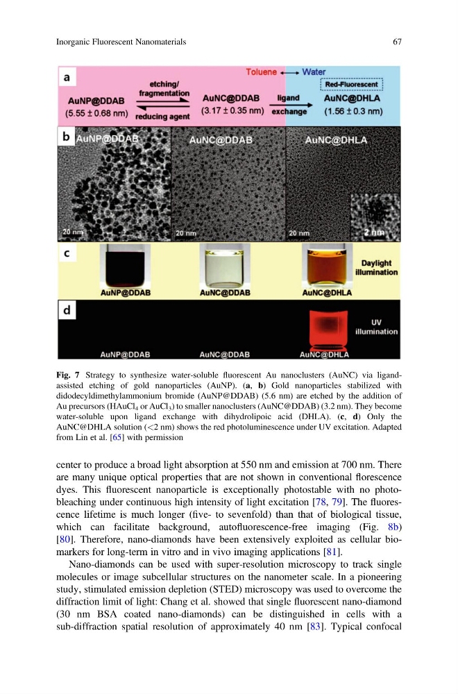
Fig. 7 Strategy to synthesize water-soluble fluorescent Au nanoclusters (AuNC) via ligandassisted etching of gold nanoparticles (AuNP). (a, b) Gold nanoparticles stabilized with
didodecyldimethylammonium bromide (AuNP@DDAB) (5.6 nm) are etched by the addition of
Au precursors (HAuCl 4 or AuCl 3 ) to smaller nanoclusters (AuNC@DDAB) (3.2 nm). They become
water-soluble upon ligand exchange with dihydrolipoic acid (DHLA). (c, d) Only the
AuNC@DHLA solution (<2 nm) shows the red photoluminescence under UV excitation. Adapted
from Lin et al. [65] with permission
Inorganic Fluorescent Nanomaterials
67
are many unique optical properties that are not shown in conventional florescence
dyes. This fluorescent nanoparticle is exceptionally photostable with no photobleaching under continuous high intensity of light excitation [78, 79]. The fluorescence lifetime is much longer (five- to sevenfold) than that of biological tissue,
which can facilitate background, autofluorescence-free imaging (Fig. 8b)
[80]. Therefore, nano-diamonds have been extensively exploited as cellular biomarkers for long-term in vitro and in vivo imaging applications [81].
Nano-diamonds can be used with super-resolution microscopy to track single
molecules or image subcellular structures on the nanometer scale. In a pioneering
study, stimulated emission depletion (STED) microscopy was used to overcome the
diffraction limit of light: Chang et al. showed that single fluorescent nano-diamond
(30 nm BSA coated nano-diamonds) can be distinguished in cells with a
sub-diffraction spatial resolution of approximately 40 nm [83]. Typical confocal
Fig. 7 Strategy to synthesize water-soluble fluorescent Au nanoclusters (AuNC) via ligandassisted etching of gold nanoparticles (AuNP). (a, b) Gold nanoparticles stabilized with
didodecyldimethylammonium bromide (AuNP@DDAB) (5.6 nm) are etched by the addition of
Au precursors (HAuCl 4 or AuCl 3 ) to smaller nanoclusters (AuNC@DDAB) (3.2 nm). They become
water-soluble upon ligand exchange with dihydrolipoic acid (DHLA). (c, d) Only the
AuNC@DHLA solution (<2 nm) shows the red photoluminescence under UV excitation. Adapted
from Lin et al. [65] with permission
Inorganic Fluorescent Nanomaterials
67