

penetration depth and prolonged observation time. 6-(benzo[d]thiazol-2
0 -yl)-2-(N,
N-dimethylamino)naphthalene (BTDAN) with disulfide group was developed as
reporter and triphenylphosphonium salt as mitochondrial targeting site. This thiol
reporter system provided selectivity and low cytotoxicity without the pH
interference [99].
3.3 pH Imaging
pH measurement is important because various diseases such as cancer, immune
dysfunction, and cystic fibrosis are associated with acid/base homeostasis [100–
102]. Small molecules [103–107] and nanoparticles [108–110] have been employed
for the ratiometric determination of pH. For example, a nanoplatform was designed
by coupling of a pH-sensitive dye, cyanine HCyC-646, or a pH-insensitive dye, Cy
7, to the bacteriophage for the pH ratiometric imaging. The emission from bacteriophage probe conjugated with HCyC-646 varies with the change of pH, whereas the
signal of probe with Cy 7 remained the same. In addition, intracellular ratiometric
pH map in RAW cells was obtained by fluorescence imaging of bacteriophage
probe with HCyC-646 [111]. Also, a pH-sensitive dendritic nanoprobe using
pentaerythritol as the core was developed, demonstrating that nanoprobes incubated
at pH 4.5 presented six times higher fluorescence than that incubated at pH 7.
Fluorescent lifetime and intensity of nanoprobes for NIR fluorescent dyes could be
activated in acidic environment owing to acid-sensitive linkage formed between
fluorescent dye and dendritic scaffold. The fluorescence intensity and lifetime can
provide both physiological and molecular information. This nanosystem with
pH-sensitive linkages can be used to noninvasively detect and analyze acidic tissues
with high sensitivity and specificity [112]. Another example is a NIR fluorescence
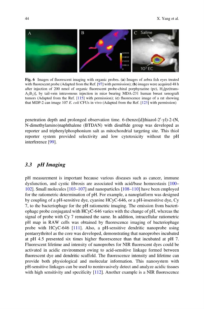
Fig. 6 Images of fluorescent imaging with organic probes. (a) Images of zebra fish eyes treated
with fluorescent probe (Adapted from the Ref. [97] with permission); (b) images were acquired 48 h
after injection of 200 nmol of organic fluorescent probe-chiral porphyrazine (pz), H 2 [pz(transA 2 B 2 )], by tail-vein intravenous injection in mice bearing MDA-231 human breast xenograft
tumors (Adapted from the Ref. [115] with permission); (c) fluorescence image of a rat showing
that MDP-2 can image 107 E. coli CFUs in vivo (Adapted from the Ref. [123] with permission)
44
X. Yang et al.
0 -yl)-2-(N,
N-dimethylamino)naphthalene (BTDAN) with disulfide group was developed as
reporter and triphenylphosphonium salt as mitochondrial targeting site. This thiol
reporter system provided selectivity and low cytotoxicity without the pH
interference [99].
3.3 pH Imaging
pH measurement is important because various diseases such as cancer, immune
dysfunction, and cystic fibrosis are associated with acid/base homeostasis [100–
102]. Small molecules [103–107] and nanoparticles [108–110] have been employed
for the ratiometric determination of pH. For example, a nanoplatform was designed
by coupling of a pH-sensitive dye, cyanine HCyC-646, or a pH-insensitive dye, Cy
7, to the bacteriophage for the pH ratiometric imaging. The emission from bacteriophage probe conjugated with HCyC-646 varies with the change of pH, whereas the
signal of probe with Cy 7 remained the same. In addition, intracellular ratiometric
pH map in RAW cells was obtained by fluorescence imaging of bacteriophage
probe with HCyC-646 [111]. Also, a pH-sensitive dendritic nanoprobe using
pentaerythritol as the core was developed, demonstrating that nanoprobes incubated
at pH 4.5 presented six times higher fluorescence than that incubated at pH 7.
Fluorescent lifetime and intensity of nanoprobes for NIR fluorescent dyes could be
activated in acidic environment owing to acid-sensitive linkage formed between
fluorescent dye and dendritic scaffold. The fluorescence intensity and lifetime can
provide both physiological and molecular information. This nanosystem with
pH-sensitive linkages can be used to noninvasively detect and analyze acidic tissues
with high sensitivity and specificity [112]. Another example is a NIR fluorescence
Fig. 6 Images of fluorescent imaging with organic probes. (a) Images of zebra fish eyes treated
with fluorescent probe (Adapted from the Ref. [97] with permission); (b) images were acquired 48 h
after injection of 200 nmol of organic fluorescent probe-chiral porphyrazine (pz), H 2 [pz(transA 2 B 2 )], by tail-vein intravenous injection in mice bearing MDA-231 human breast xenograft
tumors (Adapted from the Ref. [115] with permission); (c) fluorescence image of a rat showing
that MDP-2 can image 107 E. coli CFUs in vivo (Adapted from the Ref. [123] with permission)
44
X. Yang et al.