

3.2 Thiol Detection
Bio-thiols such as cysteine (Cys), homocysteine (Hcy), and glutathione (GSH) as
necessary reactive sulfur species play an indispensable role in cellular activity [92–
94]. For example, GSH can regulate the cellular redox activities, signal transduction,
and gene expression [95], whereas Cys and Hcy are the precursors of hydrogen
sulfide. Many diseases including cancers can be possibly caused by the abnormally
high levels of intracellular Cys [96]. Elevated amount of homocysteine might give
rise to Alzheimer’s disease or inflammatory bowel diseases. Therefore, accurate
detection of intracellular thiols is intriguing for disease diagnosis. A fluoresceinbased fluorescent probe was synthesized by conjugation of 2-cyclopentenone to
fluorescein-monoaldehyde in the presence of imidazole in THF for the detection of
various thiol-containing analytes including Cys, Hcy, GSH, Gly, Phe, Ser, Glu, Lys,
Arg, His, Ala, Gln, Met, Tyr, and cysteine. This probe itself is nonfluorescent, but
after addition of Cys, Hcy, and GSH, stronger fluorescence intensity and greater
UV-Vis spectral changes occurred because of the formation of thioether. The probe
itself is nonfluorescent without GSH, but after addition of GSH under neutral and
basic conditions, a new emission peak was observed, and the fluorescence intensity
was increased by 61-fold. This probe was also applied to monitor thiols in cells and
independent tissues and organs of zebra fish. Strong fluorescence was observed
from murine P19 embryonic carcinoma cells and 3-day-old zebra fish after incubation
with 20 μM of the probe [97] (see Fig. 6a). Another example is 4-aminonaphthalimide
dimer connected by a disulfide linker that can detect thiol quantitatively. Once thiol
triggers the cleavage of the disulfide group, 4-aminonaphthalimide is released, and its
fluorescence can be restored showing jade green color seen by naked eyes. This probe
was also applied successfully for the thiol imaging in living HeLa cells without pH
interference [98]. Also, two-photon fluorescent probe could be used to detect
the thiols in mitochondria in live cells and living tissues at 90–190 μm depth.
Compared to one-photon microscopy, two-photon microscopy can provide deeper
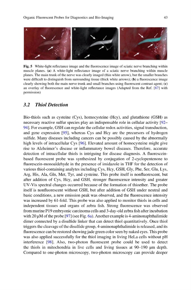
Fig. 5 White-light reflectance image and the fluorescence image of sciatic nerve branching within
muscle planes. (a) A white-light reflectance image of a sciatic nerve branching within muscle
planes. The main trunk of the nerve was clearly imaged (thin white arrow), but the smaller branches
were difficult to distinguish from surrounding tissue (thick white arrows); (b) a fluorescence image
clearly showing both the main nerve trunk and small branches using fluorescent contrast agent; (c)
an overlay of fluorescence and white-light reflectance images (Adapted from the Ref. [87] with
permission)
Organic Fluorescent Probes for Diagnostics and Bio-Imaging
43
Bio-thiols such as cysteine (Cys), homocysteine (Hcy), and glutathione (GSH) as
necessary reactive sulfur species play an indispensable role in cellular activity [92–
94]. For example, GSH can regulate the cellular redox activities, signal transduction,
and gene expression [95], whereas Cys and Hcy are the precursors of hydrogen
sulfide. Many diseases including cancers can be possibly caused by the abnormally
high levels of intracellular Cys [96]. Elevated amount of homocysteine might give
rise to Alzheimer’s disease or inflammatory bowel diseases. Therefore, accurate
detection of intracellular thiols is intriguing for disease diagnosis. A fluoresceinbased fluorescent probe was synthesized by conjugation of 2-cyclopentenone to
fluorescein-monoaldehyde in the presence of imidazole in THF for the detection of
various thiol-containing analytes including Cys, Hcy, GSH, Gly, Phe, Ser, Glu, Lys,
Arg, His, Ala, Gln, Met, Tyr, and cysteine. This probe itself is nonfluorescent, but
after addition of Cys, Hcy, and GSH, stronger fluorescence intensity and greater
UV-Vis spectral changes occurred because of the formation of thioether. The probe
itself is nonfluorescent without GSH, but after addition of GSH under neutral and
basic conditions, a new emission peak was observed, and the fluorescence intensity
was increased by 61-fold. This probe was also applied to monitor thiols in cells and
independent tissues and organs of zebra fish. Strong fluorescence was observed
from murine P19 embryonic carcinoma cells and 3-day-old zebra fish after incubation
with 20 μM of the probe [97] (see Fig. 6a). Another example is 4-aminonaphthalimide
dimer connected by a disulfide linker that can detect thiol quantitatively. Once thiol
triggers the cleavage of the disulfide group, 4-aminonaphthalimide is released, and its
fluorescence can be restored showing jade green color seen by naked eyes. This probe
was also applied successfully for the thiol imaging in living HeLa cells without pH
interference [98]. Also, two-photon fluorescent probe could be used to detect
the thiols in mitochondria in live cells and living tissues at 90–190 μm depth.
Compared to one-photon microscopy, two-photon microscopy can provide deeper
Fig. 5 White-light reflectance image and the fluorescence image of sciatic nerve branching within
muscle planes. (a) A white-light reflectance image of a sciatic nerve branching within muscle
planes. The main trunk of the nerve was clearly imaged (thin white arrow), but the smaller branches
were difficult to distinguish from surrounding tissue (thick white arrows); (b) a fluorescence image
clearly showing both the main nerve trunk and small branches using fluorescent contrast agent; (c)
an overlay of fluorescence and white-light reflectance images (Adapted from the Ref. [87] with
permission)
Organic Fluorescent Probes for Diagnostics and Bio-Imaging
43