

5-ALA was administered 3 h prior to anesthesia, and resection was conducted until
all fluorescence was completely removed while preserving neurological function.
Consequently, high rate of CRET and GTR was achieved [27]. However, increasing
resection toward the abnormality border might lead to neurological defects such as
brain tissue injury or vascular injury. In addition, 5-ALA was used for imagingguided surgery of high-grade gliomas in eloquent areas. For example, in one study,
assisted by 5-ALA fluorescence and monitoring, gross total resection (GTR) > 98%
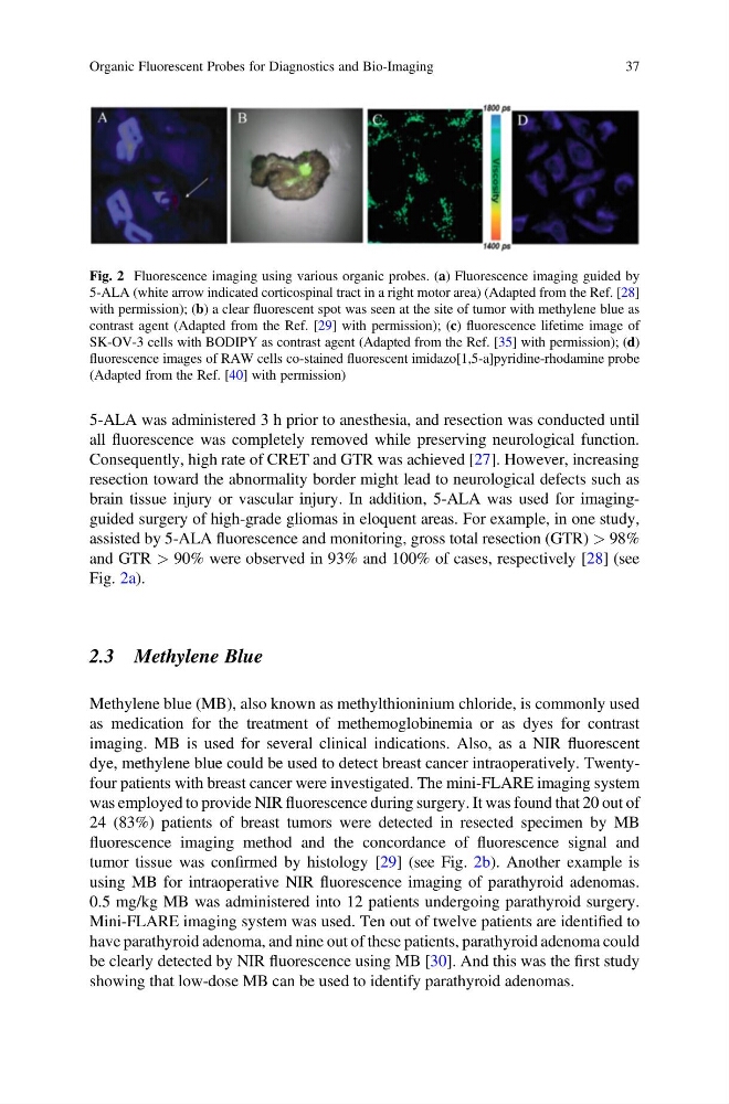
and GTR > 90% were observed in 93% and 100% of cases, respectively [28] (see
Fig. 2a).
2.3 Methylene Blue
Methylene blue (MB), also known as methylthioninium chloride, is commonly used
as medication for the treatment of methemoglobinemia or as dyes for contrast
imaging. MB is used for several clinical indications. Also, as a NIR fluorescent
dye, methylene blue could be used to detect breast cancer intraoperatively. Twentyfour patients with breast cancer were investigated. The mini-FLARE imaging system
was employed to provide NIR fluorescence during surgery. It was found that 20 out of
24 (83%) patients of breast tumors were detected in resected specimen by MB
fluorescence imaging method and the concordance of fluorescence signal and
tumor tissue was confirmed by histology [29] (see Fig. 2b). Another example is
using MB for intraoperative NIR fluorescence imaging of parathyroid adenomas.
0.5 mg/kg MB was administered into 12 patients undergoing parathyroid surgery.
Mini-FLARE imaging system was used. Ten out of twelve patients are identified to
have parathyroid adenoma, and nine out of these patients, parathyroid adenoma could
be clearly detected by NIR fluorescence using MB [30]. And this was the first study
showing that low-dose MB can be used to identify parathyroid adenomas.
Fig. 2 Fluorescence imaging using various organic probes. (a) Fluorescence imaging guided by
5-ALA (white arrow indicated corticospinal tract in a right motor area) (Adapted from the Ref. [28]
with permission); (b) a clear fluorescent spot was seen at the site of tumor with methylene blue as
contrast agent (Adapted from the Ref. [29] with permission); (c) fluorescence lifetime image of
SK-OV-3 cells with BODIPY as contrast agent (Adapted from the Ref. [35] with permission); (d)
fluorescence images of RAW cells co-stained fluorescent imidazo[1,5-a]pyridine-rhodamine probe
(Adapted from the Ref. [40] with permission)
Organic Fluorescent Probes for Diagnostics and Bio-Imaging
37
all fluorescence was completely removed while preserving neurological function.
Consequently, high rate of CRET and GTR was achieved [27]. However, increasing
resection toward the abnormality border might lead to neurological defects such as
brain tissue injury or vascular injury. In addition, 5-ALA was used for imagingguided surgery of high-grade gliomas in eloquent areas. For example, in one study,
assisted by 5-ALA fluorescence and monitoring, gross total resection (GTR) > 98%
and GTR > 90% were observed in 93% and 100% of cases, respectively [28] (see
Fig. 2a).
2.3 Methylene Blue
Methylene blue (MB), also known as methylthioninium chloride, is commonly used
as medication for the treatment of methemoglobinemia or as dyes for contrast
imaging. MB is used for several clinical indications. Also, as a NIR fluorescent
dye, methylene blue could be used to detect breast cancer intraoperatively. Twentyfour patients with breast cancer were investigated. The mini-FLARE imaging system
was employed to provide NIR fluorescence during surgery. It was found that 20 out of
24 (83%) patients of breast tumors were detected in resected specimen by MB
fluorescence imaging method and the concordance of fluorescence signal and
tumor tissue was confirmed by histology [29] (see Fig. 2b). Another example is
using MB for intraoperative NIR fluorescence imaging of parathyroid adenomas.
0.5 mg/kg MB was administered into 12 patients undergoing parathyroid surgery.
Mini-FLARE imaging system was used. Ten out of twelve patients are identified to
have parathyroid adenoma, and nine out of these patients, parathyroid adenoma could
be clearly detected by NIR fluorescence using MB [30]. And this was the first study
showing that low-dose MB can be used to identify parathyroid adenomas.
Fig. 2 Fluorescence imaging using various organic probes. (a) Fluorescence imaging guided by
5-ALA (white arrow indicated corticospinal tract in a right motor area) (Adapted from the Ref. [28]
with permission); (b) a clear fluorescent spot was seen at the site of tumor with methylene blue as
contrast agent (Adapted from the Ref. [29] with permission); (c) fluorescence lifetime image of
SK-OV-3 cells with BODIPY as contrast agent (Adapted from the Ref. [35] with permission); (d)
fluorescence images of RAW cells co-stained fluorescent imidazo[1,5-a]pyridine-rhodamine probe
(Adapted from the Ref. [40] with permission)
Organic Fluorescent Probes for Diagnostics and Bio-Imaging
37