

it takes from minutes to hours for the chromophore to mature and produce fluorescent signal, which limits their applications in real-time detection of protein-protein
interactions [86]. Apart from this, the split FP assembly is irreversible in most cases,
although there have been reports of partial recovery of split FP [89]. On the other
hand, the BiFC system achieves higher sensitivity and detects even weak interactions as it accumulates signal over time, thus prevailing over FRET sensors in this
aspect [90]. Numerous variants of split fluorescent protein-based sensors with
different spectral properties have emerged over the years, e.g., blue (EBFP), cyan
(ECFP, Cerulean, SCFP3A), green (EGFP), and yellow (EYFP, Venus, Citrine)
mutants of Aequorea victoria GFP [91–94]. In addition split variants of red and
far-red FPs such as mRFP1 [94], mCherry [95], DsRed-monomer [96], and mKate
[97] have their origin from other fluorescent proteins. As a result of common origin
from same FP, fragments from different color mutants with complementary fragments also yield cross-associated species with distinct spectral properties, which
extends the application of these systems in competitive protein binding interactions
[92, 98]. The other possibility of using two split FPs of different origins capable of
hybrid formation may be applied to visualization of two independent pairs of
protein-protein interactions [95, 96]. The combination of cross-associated BiFC
and non-cross-associated BiFC system enables simultaneous detection of three
pairs of protein-protein interactions taking place within the same cells at any given
time point [97].
In the cases of certain BiFC, chimeric proteins consisting of fragments from
different proteins sometimes reconstitute chromophore and emit fluorescence, which
offers diverse fluorescent shades capable of tracking multiple events simultaneously
in cells [96]. As an example of such a multicolor BiFC chimera, the event of liganddependent oligomerization (homodimer and heterodimer) between adenosine A 2A
and dopamine D2 receptors was evaluated effectively in a differentiated neuronal
cell model [99].
Another emerging strategy is based on incorporation of unnatural amino acids
(UAAs) into a natural chromophore of fluorescent protein for developing single FP
sensors. The incorporation UAA to proteins has recently emerged as a strategy to
generate novel rationally engineered single FP sensors [100]. One of the earliest
reports on this approach was made by Yun and coworkers wherein GFP-dopa mutant
was generated by replacing all tyrosine residues in the GFP with metal-chelating
L-DOPA [101]. The mutant variant functioned as a selective Cu
2+ sensor. Similarly
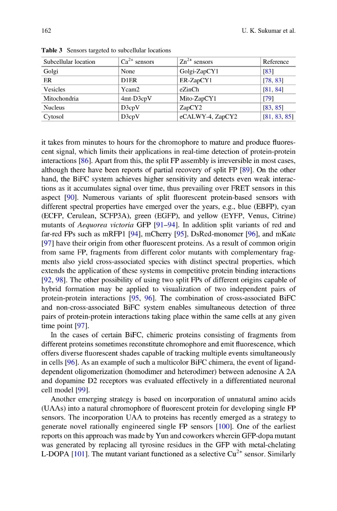
Table 3 Sensors targeted to subcellular locations
Subcellular location
Ca
2+ sensors
Zn
2+ sensors
Reference
Golgi
None
Golgi-ZapCY1
[83]
ER
D1ER
ER-ZapCY1
[78, 83]
Vesicles
Ycam2
eZinCh
[81, 84]
Mitochondria
4mt-D3cpV
Mito-ZapCY1
[79]
Nucleus
D3cpV
ZapCY2
[83, 85]
Cytosol
D3cpV
eCALWY-4, ZapCY2
[81, 83, 85]
162
U. K. Sukumar et al.
interactions [86]. Apart from this, the split FP assembly is irreversible in most cases,
although there have been reports of partial recovery of split FP [89]. On the other
hand, the BiFC system achieves higher sensitivity and detects even weak interactions as it accumulates signal over time, thus prevailing over FRET sensors in this
aspect [90]. Numerous variants of split fluorescent protein-based sensors with
different spectral properties have emerged over the years, e.g., blue (EBFP), cyan
(ECFP, Cerulean, SCFP3A), green (EGFP), and yellow (EYFP, Venus, Citrine)
mutants of Aequorea victoria GFP [91–94]. In addition split variants of red and
far-red FPs such as mRFP1 [94], mCherry [95], DsRed-monomer [96], and mKate
[97] have their origin from other fluorescent proteins. As a result of common origin
from same FP, fragments from different color mutants with complementary fragments also yield cross-associated species with distinct spectral properties, which
extends the application of these systems in competitive protein binding interactions
[92, 98]. The other possibility of using two split FPs of different origins capable of
hybrid formation may be applied to visualization of two independent pairs of
protein-protein interactions [95, 96]. The combination of cross-associated BiFC
and non-cross-associated BiFC system enables simultaneous detection of three
pairs of protein-protein interactions taking place within the same cells at any given
time point [97].
In the cases of certain BiFC, chimeric proteins consisting of fragments from
different proteins sometimes reconstitute chromophore and emit fluorescence, which
offers diverse fluorescent shades capable of tracking multiple events simultaneously
in cells [96]. As an example of such a multicolor BiFC chimera, the event of liganddependent oligomerization (homodimer and heterodimer) between adenosine A 2A
and dopamine D2 receptors was evaluated effectively in a differentiated neuronal
cell model [99].
Another emerging strategy is based on incorporation of unnatural amino acids
(UAAs) into a natural chromophore of fluorescent protein for developing single FP
sensors. The incorporation UAA to proteins has recently emerged as a strategy to
generate novel rationally engineered single FP sensors [100]. One of the earliest
reports on this approach was made by Yun and coworkers wherein GFP-dopa mutant
was generated by replacing all tyrosine residues in the GFP with metal-chelating
L-DOPA [101]. The mutant variant functioned as a selective Cu
2+ sensor. Similarly
Table 3 Sensors targeted to subcellular locations
Subcellular location
Ca
2+ sensors
Zn
2+ sensors
Reference
Golgi
None
Golgi-ZapCY1
[83]
ER
D1ER
ER-ZapCY1
[78, 83]
Vesicles
Ycam2
eZinCh
[81, 84]
Mitochondria
4mt-D3cpV
Mito-ZapCY1
[79]
Nucleus
D3cpV
ZapCY2
[83, 85]
Cytosol
D3cpV
eCALWY-4, ZapCY2
[81, 83, 85]
162
U. K. Sukumar et al.