

between target proteins, the split FP halves are brought together, resulting in
assembly of functional fluorescent protein with intact chromophore which results
in the appearance of fluorescence. This approach provides straightforward interpretation for extent and location of target protein interaction in the cell. A drawback of
BiFC as compared to FRET sensors is that upon reassociation of split FP fragments,
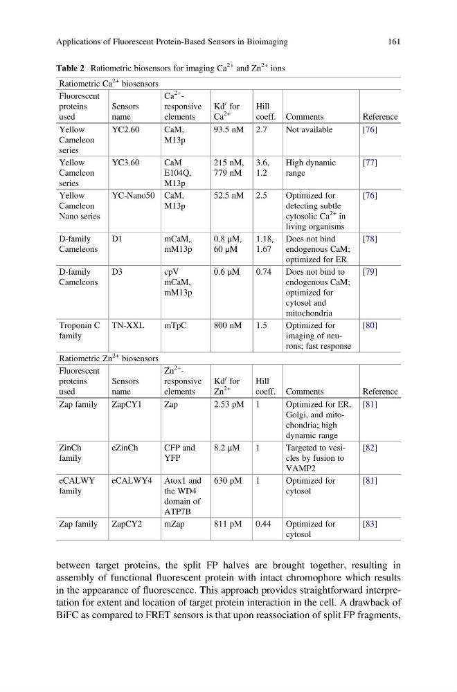
Table 2 Ratiometric biosensors for imaging Ca
2+ and Zn
2+ ions
Ratiometric Ca
2+ biosensors
Fluorescent
proteins
used
Sensors
name
Ca
2+ -
responsive
elements
Kd
0 for
Ca
2+
Hill
coeff. Comments
Reference
Yellow
Cameleon
series
YC2.60
CaM,
M13p
93.5 nM 2.7
Not available
[76]
Yellow
Cameleon
series
YC3.60
CaM
E104Q,
M13p
215 nM,
779 nM
3.6,
1.2
High dynamic
range
[77]
Yellow
Cameleon
Nano series
YC-Nano50 CaM,
M13p
52.5 nM 2.5
Optimized for
detecting subtle
cytosolic Ca
2+ in
living organisms
[76]
D-family
Cameleons
D1
mCaM,
mM13p
0.8 μM,
60 μM
1.18,
1.67
Does not bind
endogenous CaM;
optimized for ER
[78]
D-family
Cameleons
D3
cpV
mCaM,
mM13p
0.6 μM
0.74
Does not bind to
endogenous CaM;
optimized for
cytosol and
mitochondria
[79]
Troponin C
family
TN-XXL
mTpC
800 nM
1.5
Optimized for
imaging of neurons; fast response
[80]
Ratiometric Zn
2+ biosensors
Fluorescent
proteins
used
Sensors
name
Zn
2+ -
responsive
elements
Kd
0 for
Zn
2+
Hill
coeff. Comments
Reference
Zap family
ZapCY1
Zap
2.53 pM 1
Optimized for ER,
Golgi, and mitochondria; high
dynamic range
[81]
ZinCh
family
eZinCh
CFP and
YFP
8.2 μM
1
Targeted to vesicles by fusion to
VAMP2
[82]
eCALWY
family
eCALWY4 Atox1 and
the WD4
domain of
ATP7B
630 pM
1
Optimized for
cytosol
[81]
Zap family
ZapCY2
mZap
811 pM
0.44
Optimized for
cytosol
[83]
Applications of Fluorescent Protein-Based Sensors in Bioimaging
161
assembly of functional fluorescent protein with intact chromophore which results
in the appearance of fluorescence. This approach provides straightforward interpretation for extent and location of target protein interaction in the cell. A drawback of
BiFC as compared to FRET sensors is that upon reassociation of split FP fragments,
Table 2 Ratiometric biosensors for imaging Ca
2+ and Zn
2+ ions
Ratiometric Ca
2+ biosensors
Fluorescent
proteins
used
Sensors
name
Ca
2+ -
responsive
elements
Kd
0 for
Ca
2+
Hill
coeff. Comments
Reference
Yellow
Cameleon
series
YC2.60
CaM,
M13p
93.5 nM 2.7
Not available
[76]
Yellow
Cameleon
series
YC3.60
CaM
E104Q,
M13p
215 nM,
779 nM
3.6,
1.2
High dynamic
range
[77]
Yellow
Cameleon
Nano series
YC-Nano50 CaM,
M13p
52.5 nM 2.5
Optimized for
detecting subtle
cytosolic Ca
2+ in
living organisms
[76]
D-family
Cameleons
D1
mCaM,
mM13p
0.8 μM,
60 μM
1.18,
1.67
Does not bind
endogenous CaM;
optimized for ER
[78]
D-family
Cameleons
D3
cpV
mCaM,
mM13p
0.6 μM
0.74
Does not bind to
endogenous CaM;
optimized for
cytosol and
mitochondria
[79]
Troponin C
family
TN-XXL
mTpC
800 nM
1.5
Optimized for
imaging of neurons; fast response
[80]
Ratiometric Zn
2+ biosensors
Fluorescent
proteins
used
Sensors
name
Zn
2+ -
responsive
elements
Kd
0 for
Zn
2+
Hill
coeff. Comments
Reference
Zap family
ZapCY1
Zap
2.53 pM 1
Optimized for ER,
Golgi, and mitochondria; high
dynamic range
[81]
ZinCh
family
eZinCh
CFP and
YFP
8.2 μM
1
Targeted to vesicles by fusion to
VAMP2
[82]
eCALWY
family
eCALWY4 Atox1 and
the WD4
domain of
ATP7B
630 pM
1
Optimized for
cytosol
[81]
Zap family
ZapCY2
mZap
811 pM
0.44
Optimized for
cytosol
[83]
Applications of Fluorescent Protein-Based Sensors in Bioimaging
161