

3.1.1 Fluorescent Biosensors as Tool for Detection of Intracellular Ion
Concentrations
The pH-responsive fluorescent proteins have been used to monitor exocytosis and
recycling of proteins based on the rationale of prevalent acidic pH of the secretory
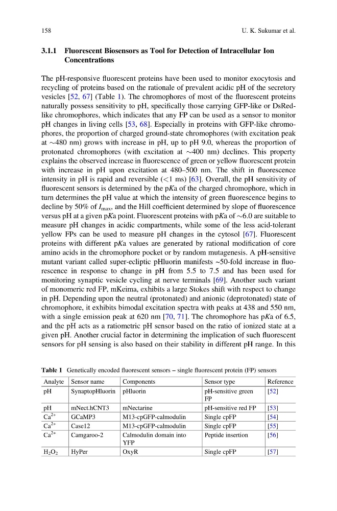
vesicles [52, 67] (Table 1). The chromophores of most of the fluorescent proteins
naturally possess sensitivity to pH, specifically those carrying GFP-like or DsRedlike chromophores, which indicates that any FP can be used as a sensor to monitor
pH changes in living cells [53, 68]. Especially in proteins with GFP-like chromophores, the proportion of charged ground-state chromophores (with excitation peak
at $480 nm) grows with increase in pH, up to pH 9.0, whereas the proportion of
protonated chromophores (with excitation at $400 nm) declines. This property
explains the observed increase in fluorescence of green or yellow fluorescent protein
with increase in pH upon excitation at 480–500 nm. The shift in fluorescence
intensity in pH is rapid and reversible (<1 ms) [63]. Overall, the pH sensitivity of
fluorescent sensors is determined by the pKa of the charged chromophore, which in
turn determines the pH value at which the intensity of green fluorescence begins to
decline by 50% of I max , and the Hill coefficient determined by slope of fluorescence
versus pH at a given pKa point. Fluorescent proteins with pKa of $6.0 are suitable to
measure pH changes in acidic compartments, while some of the less acid-tolerant
yellow FPs can be used to measure pH changes in the cytosol [67]. Fluorescent
proteins with different pKa values are generated by rational modification of core
amino acids in the chromophore pocket or by random mutagenesis. A pH-sensitive
mutant variant called super-ecliptic pHluorin manifests ~50-fold increase in fluorescence in response to change in pH from 5.5 to 7.5 and has been used for
monitoring synaptic vesicle cycling at nerve terminals [69]. Another such variant
of monomeric red FP, mKeima, exhibits a large Stokes shift with respect to change
in pH. Depending upon the neutral (protonated) and anionic (deprotonated) state of
chromophore, it exhibits bimodal excitation spectra with peaks at 438 and 550 nm,
with a single emission peak at 620 nm [70, 71]. The chromophore has pKa of 6.5,
and the pH acts as a ratiometric pH sensor based on the ratio of ionized state at a
given pH. Another crucial factor in determining the implication of such fluorescent
sensors for pH sensing is also based on their stability in different pH range. In this
Table 1 Genetically encoded fluorescent sensors – single fluorescent protein (FP) sensors
Analyte Sensor name
Components
Sensor type
Reference
pH
SynaptopHluorin pHluorin
pH-sensitive green
FP
[52]
pH
mNect.hCNT3
mNectarine
pH-sensitive red FP
[53]
Ca
2+
GCaMP3
M13-cpGFP-calmodulin
Single cpFP
[54]
Ca
2+
Case12
M13-cpGFP-calmodulin
Single cpFP
[55]
Ca
2+
Camgaroo-2
Calmodulin domain into
YFP
Peptide insertion
[56]
H 2 O 2
HyPer
OxyR
Single cpFP
[57]
158
U. K. Sukumar et al.
Concentrations
The pH-responsive fluorescent proteins have been used to monitor exocytosis and
recycling of proteins based on the rationale of prevalent acidic pH of the secretory
vesicles [52, 67] (Table 1). The chromophores of most of the fluorescent proteins
naturally possess sensitivity to pH, specifically those carrying GFP-like or DsRedlike chromophores, which indicates that any FP can be used as a sensor to monitor
pH changes in living cells [53, 68]. Especially in proteins with GFP-like chromophores, the proportion of charged ground-state chromophores (with excitation peak
at $480 nm) grows with increase in pH, up to pH 9.0, whereas the proportion of
protonated chromophores (with excitation at $400 nm) declines. This property
explains the observed increase in fluorescence of green or yellow fluorescent protein
with increase in pH upon excitation at 480–500 nm. The shift in fluorescence
intensity in pH is rapid and reversible (<1 ms) [63]. Overall, the pH sensitivity of
fluorescent sensors is determined by the pKa of the charged chromophore, which in
turn determines the pH value at which the intensity of green fluorescence begins to
decline by 50% of I max , and the Hill coefficient determined by slope of fluorescence
versus pH at a given pKa point. Fluorescent proteins with pKa of $6.0 are suitable to
measure pH changes in acidic compartments, while some of the less acid-tolerant
yellow FPs can be used to measure pH changes in the cytosol [67]. Fluorescent
proteins with different pKa values are generated by rational modification of core
amino acids in the chromophore pocket or by random mutagenesis. A pH-sensitive
mutant variant called super-ecliptic pHluorin manifests ~50-fold increase in fluorescence in response to change in pH from 5.5 to 7.5 and has been used for
monitoring synaptic vesicle cycling at nerve terminals [69]. Another such variant
of monomeric red FP, mKeima, exhibits a large Stokes shift with respect to change
in pH. Depending upon the neutral (protonated) and anionic (deprotonated) state of
chromophore, it exhibits bimodal excitation spectra with peaks at 438 and 550 nm,
with a single emission peak at 620 nm [70, 71]. The chromophore has pKa of 6.5,
and the pH acts as a ratiometric pH sensor based on the ratio of ionized state at a
given pH. Another crucial factor in determining the implication of such fluorescent
sensors for pH sensing is also based on their stability in different pH range. In this
Table 1 Genetically encoded fluorescent sensors – single fluorescent protein (FP) sensors
Analyte Sensor name
Components
Sensor type
Reference
pH
SynaptopHluorin pHluorin
pH-sensitive green
FP
[52]
pH
mNect.hCNT3
mNectarine
pH-sensitive red FP
[53]
Ca
2+
GCaMP3
M13-cpGFP-calmodulin
Single cpFP
[54]
Ca
2+
Case12
M13-cpGFP-calmodulin
Single cpFP
[55]
Ca
2+
Camgaroo-2
Calmodulin domain into
YFP
Peptide insertion
[56]
H 2 O 2
HyPer
OxyR
Single cpFP
[57]
158
U. K. Sukumar et al.