

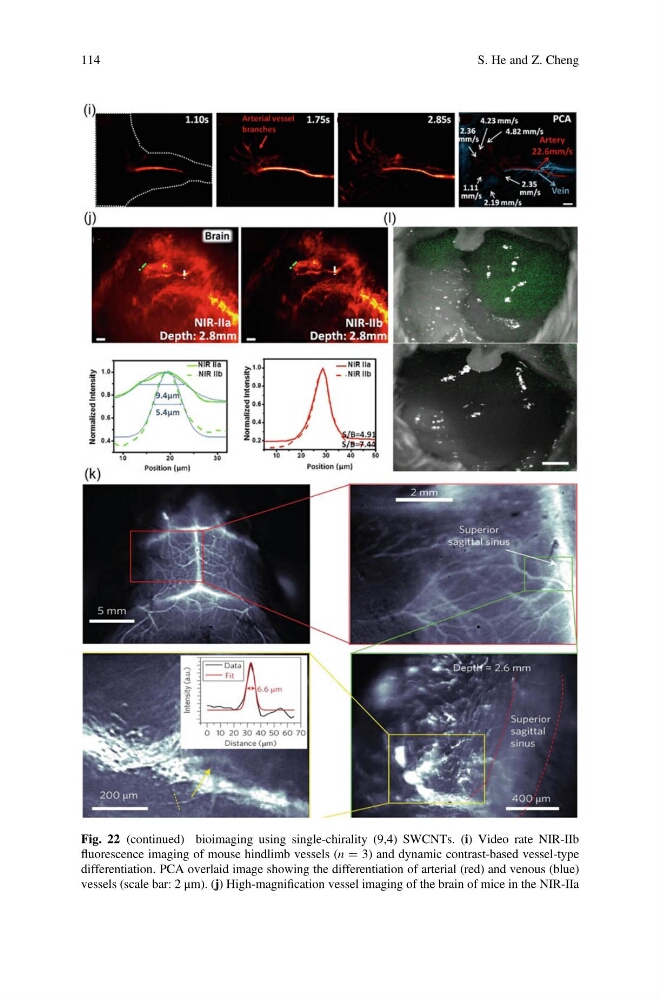
Fig. 22 (continued) bioimaging using single-chirality (9,4) SWCNTs. (i) Video rate NIR-IIb
fluorescence imaging of mouse hindlimb vessels (n ¼ 3) and dynamic contrast-based vessel-type
differentiation. PCA overlaid image showing the differentiation of arterial (red) and venous (blue)
vessels (scale bar: 2 μm). (j) High-magnification vessel imaging of the brain of mice in the NIR-IIa
114
S. He and Z. Cheng
fluorescence imaging of mouse hindlimb vessels (n ¼ 3) and dynamic contrast-based vessel-type
differentiation. PCA overlaid image showing the differentiation of arterial (red) and venous (blue)
vessels (scale bar: 2 μm). (j) High-magnification vessel imaging of the brain of mice in the NIR-IIa
114
S. He and Z. Cheng