

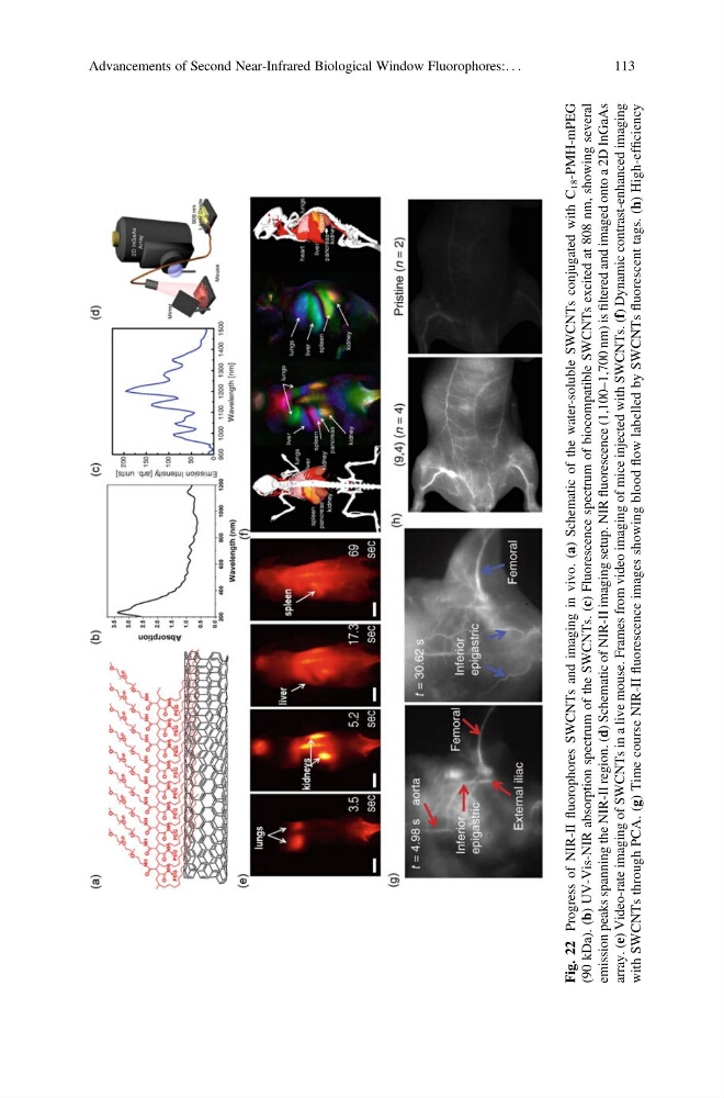
Fig. 22
Progress of NIR-II
fluorophores SWCNTs and imaging in vivo. (a) Schematic of the water-soluble SWCNTs conjugated with C
18 -PMH-mPEG
(90 kDa). (b) UV-Vis-NIR absorption spectrum of the SWCNTs. (c) Fluorescence spectrum of biocompatible SWCNTs excited at 808 nm, showing several
emission peaks spanning the NIR-II region. (d) Schematic of NIR-II imaging setup. NIR
fluorescence (1,100–1,700 nm) is
filtered and imaged onto a 2D InGaAs
array. (e) Video-rate imaging of SWCNTs in a live mouse. Frames from video imaging of mice injected with SWCNTs. (f) Dynamic contrast-enhanced imaging
with SWCNTs through PCA. (g) Time course NIR-II
fluorescence images showing blood
flow labelled by SWCNTs
fluorescent tags. (h) High-efficiency
Advancements of Second Near-Infrared Biological Window Fluorophores:. . .
113
Progress of NIR-II
fluorophores SWCNTs and imaging in vivo. (a) Schematic of the water-soluble SWCNTs conjugated with C
18 -PMH-mPEG
(90 kDa). (b) UV-Vis-NIR absorption spectrum of the SWCNTs. (c) Fluorescence spectrum of biocompatible SWCNTs excited at 808 nm, showing several
emission peaks spanning the NIR-II region. (d) Schematic of NIR-II imaging setup. NIR
fluorescence (1,100–1,700 nm) is
filtered and imaged onto a 2D InGaAs
array. (e) Video-rate imaging of SWCNTs in a live mouse. Frames from video imaging of mice injected with SWCNTs. (f) Dynamic contrast-enhanced imaging
with SWCNTs through PCA. (g) Time course NIR-II
fluorescence images showing blood
flow labelled by SWCNTs
fluorescent tags. (h) High-efficiency
Advancements of Second Near-Infrared Biological Window Fluorophores:. . .
113