

1 Introduction
Fluorescence molecular imaging (FMI) utilizes molecular probes to label target
organisms. Under certain external conditions, the molecular probe releases a fluorescent light in the visible or near-infrared spectrum using high-sensitivity detection
equipment for fluorescence. The signal is collected, and the position and intensity of
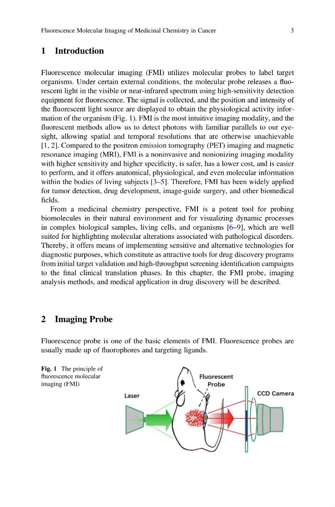
the fluorescent light source are displayed to obtain the physiological activity information of the organism (Fig. 1). FMI is the most intuitive imaging modality, and the
fluorescent methods allow us to detect photons with familiar parallels to our eyesight, allowing spatial and temporal resolutions that are otherwise unachievable
[1, 2]. Compared to the positron emission tomography (PET) imaging and magnetic
resonance imaging (MRI), FMI is a noninvasive and nonionizing imaging modality
with higher sensitivity and higher specificity, is safer, has a lower cost, and is easier
to perform, and it offers anatomical, physiological, and even molecular information
within the bodies of living subjects [3–5]. Therefore, FMI has been widely applied
for tumor detection, drug development, image-guide surgery, and other biomedical
fields.
From a medicinal chemistry perspective, FMI is a potent tool for probing
biomolecules in their natural environment and for visualizing dynamic processes
in complex biological samples, living cells, and organisms [6–9], which are well
suited for highlighting molecular alterations associated with pathological disorders.
Thereby, it offers means of implementing sensitive and alternative technologies for
diagnostic purposes, which constitute as attractive tools for drug discovery programs
from initial target validation and high-throughput screening identification campaigns
to the final clinical translation phases. In this chapter, the FMI probe, imaging
analysis methods, and medical application in drug discovery will be described.
2 Imaging Probe
Fluorescence probe is one of the basic elements of FMI. Fluorescence probes are
usually made up of fluorophores and targeting ligands.
Fig. 1 The principle of
fluorescence molecular
imaging (FMI)
Fluorescence Molecular Imaging of Medicinal Chemistry in Cancer
3
Fluorescence molecular imaging (FMI) utilizes molecular probes to label target
organisms. Under certain external conditions, the molecular probe releases a fluorescent light in the visible or near-infrared spectrum using high-sensitivity detection
equipment for fluorescence. The signal is collected, and the position and intensity of
the fluorescent light source are displayed to obtain the physiological activity information of the organism (Fig. 1). FMI is the most intuitive imaging modality, and the
fluorescent methods allow us to detect photons with familiar parallels to our eyesight, allowing spatial and temporal resolutions that are otherwise unachievable
[1, 2]. Compared to the positron emission tomography (PET) imaging and magnetic
resonance imaging (MRI), FMI is a noninvasive and nonionizing imaging modality
with higher sensitivity and higher specificity, is safer, has a lower cost, and is easier
to perform, and it offers anatomical, physiological, and even molecular information
within the bodies of living subjects [3–5]. Therefore, FMI has been widely applied
for tumor detection, drug development, image-guide surgery, and other biomedical
fields.
From a medicinal chemistry perspective, FMI is a potent tool for probing
biomolecules in their natural environment and for visualizing dynamic processes
in complex biological samples, living cells, and organisms [6–9], which are well
suited for highlighting molecular alterations associated with pathological disorders.
Thereby, it offers means of implementing sensitive and alternative technologies for
diagnostic purposes, which constitute as attractive tools for drug discovery programs
from initial target validation and high-throughput screening identification campaigns
to the final clinical translation phases. In this chapter, the FMI probe, imaging
analysis methods, and medical application in drug discovery will be described.
2 Imaging Probe
Fluorescence probe is one of the basic elements of FMI. Fluorescence probes are
usually made up of fluorophores and targeting ligands.
Fig. 1 The principle of
fluorescence molecular
imaging (FMI)
Fluorescence Molecular Imaging of Medicinal Chemistry in Cancer
3