

because of their high photostability, broad absorption range, large extinction coefficient, and narrow, tunable emission wavelengths. In addition, among the NIR-II
emitting probes reported until now, QDs show the highest NIR-II fluorescence
quantum yields (up to 15.5%) [19].
4.1 Mechanism of NIR-II Emission
The emission wavelengths could be rationally tuned by changing the QDs’ sizes and
compositions. Since the excitons are confined in all three spatial dimensions, the
electrons of QDs are quantized to certain energies, similar to those of organic dyes.
As the confinement energy depends on the quantum dots size, both absorption onset
and fluorescence emission can be tuned by changing the size of the QDs during
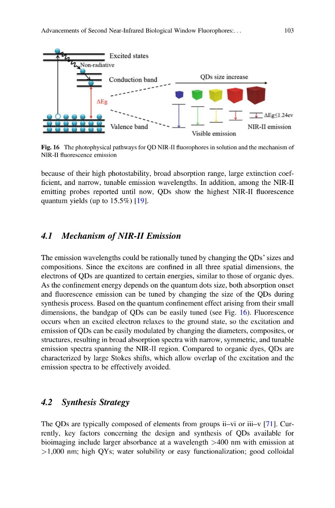
synthesis process. Based on the quantum confinement effect arising from their small
dimensions, the bandgap of QDs can be easily tuned (see Fig. 16). Fluorescence
occurs when an excited electron relaxes to the ground state, so the excitation and
emission of QDs can be easily modulated by changing the diameters, composites, or
structures, resulting in broad absorption spectra with narrow, symmetric, and tunable
emission spectra spanning the NIR-II region. Compared to organic dyes, QDs are
characterized by large Stokes shifts, which allow overlap of the excitation and the
emission spectra to be effectively avoided.
4.2 Synthesis Strategy
The QDs are typically composed of elements from groups ii–vi or iii–v [71]. Currently, key factors concerning the design and synthesis of QDs available for
bioimaging include larger absorbance at a wavelength >400 nm with emission at
>1,000 nm; high QYs; water solubility or easy functionalization; good colloidal
Fig. 16 The photophysical pathways for QD NIR-II fluorophores in solution and the mechanism of
NIR-II fluorescence emission
Advancements of Second Near-Infrared Biological Window Fluorophores:. . .
103
emitting probes reported until now, QDs show the highest NIR-II fluorescence
quantum yields (up to 15.5%) [19].
4.1 Mechanism of NIR-II Emission
The emission wavelengths could be rationally tuned by changing the QDs’ sizes and
compositions. Since the excitons are confined in all three spatial dimensions, the
electrons of QDs are quantized to certain energies, similar to those of organic dyes.
As the confinement energy depends on the quantum dots size, both absorption onset
and fluorescence emission can be tuned by changing the size of the QDs during
synthesis process. Based on the quantum confinement effect arising from their small
dimensions, the bandgap of QDs can be easily tuned (see Fig. 16). Fluorescence
occurs when an excited electron relaxes to the ground state, so the excitation and
emission of QDs can be easily modulated by changing the diameters, composites, or
structures, resulting in broad absorption spectra with narrow, symmetric, and tunable
emission spectra spanning the NIR-II region. Compared to organic dyes, QDs are
characterized by large Stokes shifts, which allow overlap of the excitation and the
emission spectra to be effectively avoided.
4.2 Synthesis Strategy
The QDs are typically composed of elements from groups ii–vi or iii–v [71]. Currently, key factors concerning the design and synthesis of QDs available for
bioimaging include larger absorbance at a wavelength >400 nm with emission at
>1,000 nm; high QYs; water solubility or easy functionalization; good colloidal
Fig. 16 The photophysical pathways for QD NIR-II fluorophores in solution and the mechanism of
NIR-II fluorescence emission
Advancements of Second Near-Infrared Biological Window Fluorophores:. . .
103