

21
The surface attached molecules marked with fluorophore are excited by this evanescent field, generating a fluorescence signal, which is then detected using a CCD
camera fitted with appropriate bandpass and longpass filters (Fig. 2.6, right).
Because of the limitation of the penetration depth of the evanescent field, only
surface-attached fluorophores are excited, allowing the analysis of heterogenous or
turbid samples. The position and intensities of the fluorescent spots express the
identity and concentration of the target sample in each lane [29, 32].
The NRL Array Biosensor was used successfully for the detection of toxins and
for multiple toxins simultaneously in multiple samples, it could also detect toxin
levels as low as 500 pg/mL and quantify the toxin concentration, and finally, it could
perform toxin assays in clinical, food, and environmental samples [29]. Furthermore,
both sandwich immunoassays for protein toxins (e.g., staphylococcal enterotoxin B
[SEB] and ricin) and competitive immunoassays for low molecular weight toxins
(e.g., trinitrotoluene and fumonisin B1) were reported. [29] A schematic of the
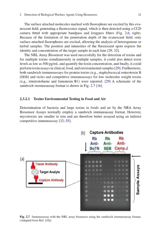
sandwich immunoassay format is shown in Fig. 2.7 [16].
2.3.2.1 Toxins Environmental Testing in Food and Air
Determination of bacteria and large toxins in foods and air by the NRA Array
Biosensor Assays normally employ a sandwich immunoassay format. However,
mycotoxins are smaller in size and are therefore better assayed using an indirect
competitive immunoassay [32–35].
Fig. 2.7 Immunoassay with the NRL array biosensor using the sandwich immunoassay format.
(Adapted from Ref. [16])
2 Detection of Biological Warfare Agents Using Biosensors
The surface attached molecules marked with fluorophore are excited by this evanescent field, generating a fluorescence signal, which is then detected using a CCD
camera fitted with appropriate bandpass and longpass filters (Fig. 2.6, right).
Because of the limitation of the penetration depth of the evanescent field, only
surface-attached fluorophores are excited, allowing the analysis of heterogenous or
turbid samples. The position and intensities of the fluorescent spots express the
identity and concentration of the target sample in each lane [29, 32].
The NRL Array Biosensor was used successfully for the detection of toxins and
for multiple toxins simultaneously in multiple samples, it could also detect toxin
levels as low as 500 pg/mL and quantify the toxin concentration, and finally, it could
perform toxin assays in clinical, food, and environmental samples [29]. Furthermore,
both sandwich immunoassays for protein toxins (e.g., staphylococcal enterotoxin B
[SEB] and ricin) and competitive immunoassays for low molecular weight toxins
(e.g., trinitrotoluene and fumonisin B1) were reported. [29] A schematic of the
sandwich immunoassay format is shown in Fig. 2.7 [16].
2.3.2.1 Toxins Environmental Testing in Food and Air
Determination of bacteria and large toxins in foods and air by the NRA Array
Biosensor Assays normally employ a sandwich immunoassay format. However,
mycotoxins are smaller in size and are therefore better assayed using an indirect
competitive immunoassay [32–35].
Fig. 2.7 Immunoassay with the NRL array biosensor using the sandwich immunoassay format.
(Adapted from Ref. [16])
2 Detection of Biological Warfare Agents Using Biosensors