

484
8 Morphogenesis
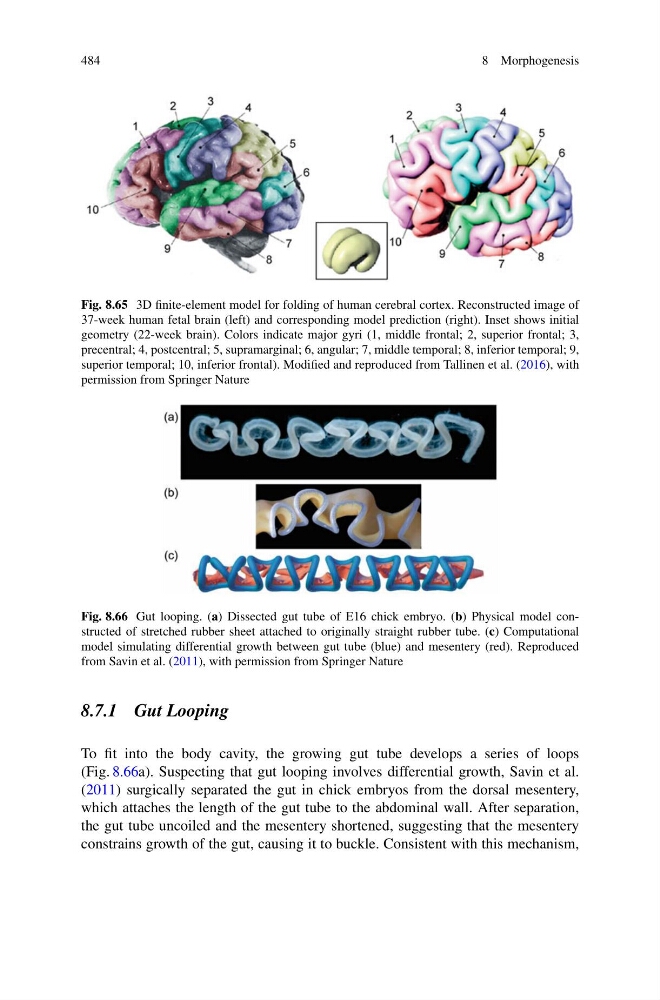
Fig. 8.65 3D finite-element model for folding of human cerebral cortex. Reconstructed image of
37-week human fetal brain (left) and corresponding model prediction (right). Inset shows initial
geometry (22-week brain). Colors indicate major gyri (1, middle frontal; 2, superior frontal; 3,
precentral; 4, postcentral; 5, supramarginal; 6, angular; 7, middle temporal; 8, inferior temporal; 9,
superior temporal; 10, inferior frontal). Modified and reproduced from Tallinen et al. (2016), with
permission from Springer Nature
Fig. 8.66 Gut looping. (a) Dissected gut tube of E16 chick embryo. (b) Physical model constructed of stretched rubber sheet attached to originally straight rubber tube. (c) Computational
model simulating differential growth between gut tube (blue) and mesentery (red). Reproduced
from Savin et al. (2011), with permission from Springer Nature
8.7.1 Gut Looping
To fit into the body cavity, the growing gut tube develops a series of loops
(Fig. 8.66a). Suspecting that gut looping involves differential growth, Savin et al.
(2011) surgically separated the gut in chick embryos from the dorsal mesentery,
which attaches the length of the gut tube to the abdominal wall. After separation,
the gut tube uncoiled and the mesentery shortened, suggesting that the mesentery
constrains growth of the gut, causing it to buckle. Consistent with this mechanism,
8 Morphogenesis
Fig. 8.65 3D finite-element model for folding of human cerebral cortex. Reconstructed image of
37-week human fetal brain (left) and corresponding model prediction (right). Inset shows initial
geometry (22-week brain). Colors indicate major gyri (1, middle frontal; 2, superior frontal; 3,
precentral; 4, postcentral; 5, supramarginal; 6, angular; 7, middle temporal; 8, inferior temporal; 9,
superior temporal; 10, inferior frontal). Modified and reproduced from Tallinen et al. (2016), with
permission from Springer Nature
Fig. 8.66 Gut looping. (a) Dissected gut tube of E16 chick embryo. (b) Physical model constructed of stretched rubber sheet attached to originally straight rubber tube. (c) Computational
model simulating differential growth between gut tube (blue) and mesentery (red). Reproduced
from Savin et al. (2011), with permission from Springer Nature
8.7.1 Gut Looping
To fit into the body cavity, the growing gut tube develops a series of loops
(Fig. 8.66a). Suspecting that gut looping involves differential growth, Savin et al.
(2011) surgically separated the gut in chick embryos from the dorsal mesentery,
which attaches the length of the gut tube to the abdominal wall. After separation,
the gut tube uncoiled and the mesentery shortened, suggesting that the mesentery
constrains growth of the gut, causing it to buckle. Consistent with this mechanism,