

482
8 Morphogenesis
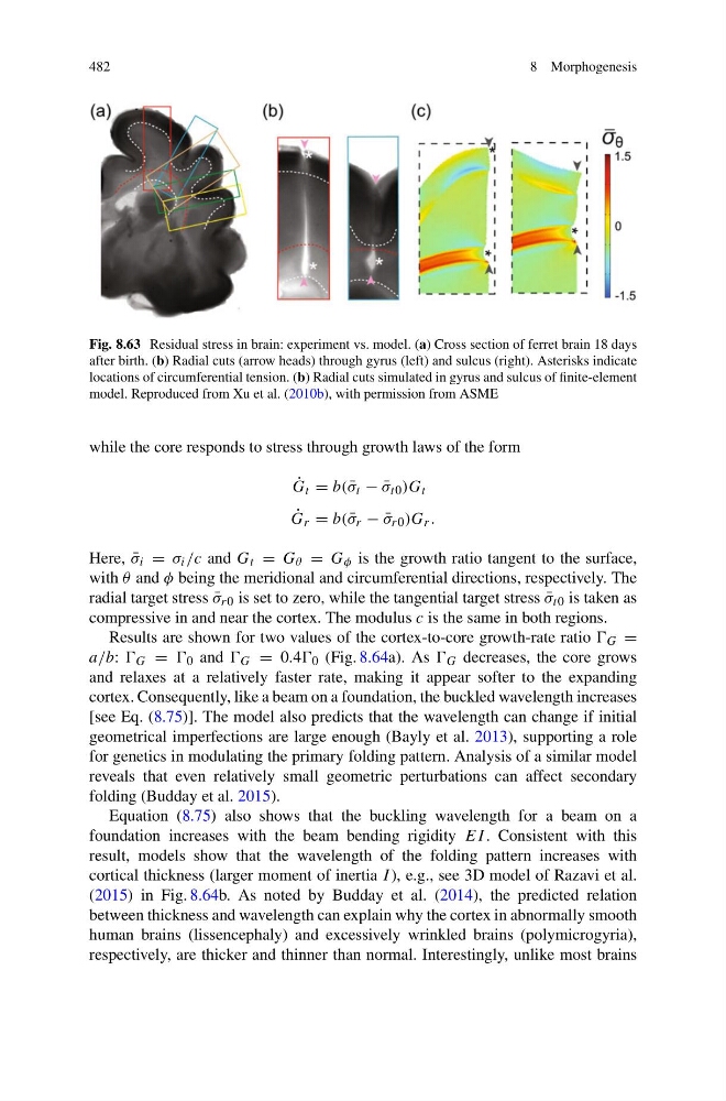
Fig. 8.63 Residual stress in brain: experiment vs. model. (a) Cross section of ferret brain 18 days
after birth. (b) Radial cuts (arrow heads) through gyrus (left) and sulcus (right). Asterisks indicate
locations of circumferential tension. (b) Radial cuts simulated in gyrus and sulcus of finite-element
model. Reproduced from Xu et al. (2010b), with permission from ASME
while the core responds to stress through growth laws of the form
˙
G t = b( ¯
σ t − ¯
σ t0 )G t
˙
G r = b( ¯
σ r − ¯
σ r0 )G r .
Here, ¯
σ i = σ i /c and G t = G θ = G φ is the growth ratio tangent to the surface,
with θ and φ being the meridional and circumferential directions, respectively. The
radial target stress ¯
σ r0 is set to zero, while the tangential target stress ¯
σ t0 is taken as
compressive in and near the cortex. The modulus c is the same in both regions.
Results are shown for two values of the cortex-to-core growth-rate ratio G =
a/b: G = 0 and G = 0.4 0 (Fig. 8.64a). As G decreases, the core grows
and relaxes at a relatively faster rate, making it appear softer to the expanding
cortex. Consequently, like a beam on a foundation, the buckled wavelength increases
[see Eq. (8.75)]. The model also predicts that the wavelength can change if initial
geometrical imperfections are large enough (Bayly et al. 2013), supporting a role
for genetics in modulating the primary folding pattern. Analysis of a similar model
reveals that even relatively small geometric perturbations can affect secondary
folding (Budday et al. 2015).
Equation (8.75) also shows that the buckling wavelength for a beam on a
foundation increases with the beam bending rigidity EI . Consistent with this
result, models show that the wavelength of the folding pattern increases with
cortical thickness (larger moment of inertia I ), e.g., see 3D model of Razavi et al.
(2015) in Fig. 8.64b. As noted by Budday et al. (2014), the predicted relation
between thickness and wavelength can explain why the cortex in abnormally smooth
human brains (lissencephaly) and excessively wrinkled brains (polymicrogyria),
respectively, are thicker and thinner than normal. Interestingly, unlike most brains
8 Morphogenesis
Fig. 8.63 Residual stress in brain: experiment vs. model. (a) Cross section of ferret brain 18 days
after birth. (b) Radial cuts (arrow heads) through gyrus (left) and sulcus (right). Asterisks indicate
locations of circumferential tension. (b) Radial cuts simulated in gyrus and sulcus of finite-element
model. Reproduced from Xu et al. (2010b), with permission from ASME
while the core responds to stress through growth laws of the form
˙
G t = b( ¯
σ t − ¯
σ t0 )G t
˙
G r = b( ¯
σ r − ¯
σ r0 )G r .
Here, ¯
σ i = σ i /c and G t = G θ = G φ is the growth ratio tangent to the surface,
with θ and φ being the meridional and circumferential directions, respectively. The
radial target stress ¯
σ r0 is set to zero, while the tangential target stress ¯
σ t0 is taken as
compressive in and near the cortex. The modulus c is the same in both regions.
Results are shown for two values of the cortex-to-core growth-rate ratio G =
a/b: G = 0 and G = 0.4 0 (Fig. 8.64a). As G decreases, the core grows
and relaxes at a relatively faster rate, making it appear softer to the expanding
cortex. Consequently, like a beam on a foundation, the buckled wavelength increases
[see Eq. (8.75)]. The model also predicts that the wavelength can change if initial
geometrical imperfections are large enough (Bayly et al. 2013), supporting a role
for genetics in modulating the primary folding pattern. Analysis of a similar model
reveals that even relatively small geometric perturbations can affect secondary
folding (Budday et al. 2015).
Equation (8.75) also shows that the buckling wavelength for a beam on a
foundation increases with the beam bending rigidity EI . Consistent with this
result, models show that the wavelength of the folding pattern increases with
cortical thickness (larger moment of inertia I ), e.g., see 3D model of Razavi et al.
(2015) in Fig. 8.64b. As noted by Budday et al. (2014), the predicted relation
between thickness and wavelength can explain why the cortex in abnormally smooth
human brains (lissencephaly) and excessively wrinkled brains (polymicrogyria),
respectively, are thicker and thinner than normal. Interestingly, unlike most brains