

8.6 Neural Development
457
Fig. 8.41 3D model for axolotl neurulation. Embryo (grayscale images) and model (colored
images) are shown for two stages of development. Reproduced from Chen and Brodland (2008),
with permission from IOP Publishing
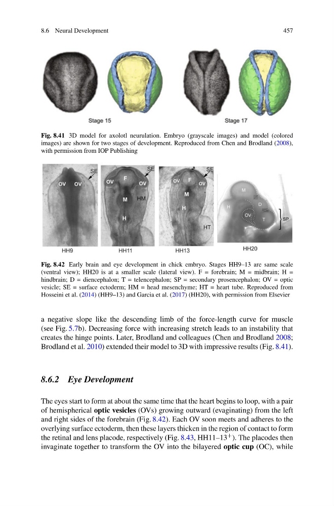
Fig. 8.42 Early brain and eye development in chick embryo. Stages HH9–13 are same scale
(ventral view); HH20 is at a smaller scale (lateral view). F = forebrain; M = midbrain; H =
hindbrain; D = diencephalon; T = telencephalon; SP = secondary prosencephalon; OV = optic
vesicle; SE = surface ectoderm; HM = head mesenchyme; HT = heart tube. Reproduced from
Hosseini et al. (2014) (HH9–13) and Garcia et al. (2017) (HH20), with permission from Elsevier
a negative slope like the descending limb of the force-length curve for muscle
(see Fig. 5.7b). Decreasing force with increasing stretch leads to an instability that
creates the hinge points. Later, Brodland and colleagues (Chen and Brodland 2008;
Brodland et al. 2010) extended their model to 3D with impressive results (Fig. 8.41).
8.6.2 Eye Development
The eyes start to form at about the same time that the heart begins to loop, with a pair
of hemispherical optic vesicles (OVs) growing outward (evaginating) from the left
and right sides of the forebrain (Fig. 8.42). Each OV soon meets and adheres to the
overlying surface ectoderm, then these layers thicken in the region of contact to form
the retinal and lens placode, respectively (Fig. 8.43, HH11–13 + ). The placodes then
invaginate together to transform the OV into the bilayered optic cup (OC), while
457
Fig. 8.41 3D model for axolotl neurulation. Embryo (grayscale images) and model (colored
images) are shown for two stages of development. Reproduced from Chen and Brodland (2008),
with permission from IOP Publishing
Fig. 8.42 Early brain and eye development in chick embryo. Stages HH9–13 are same scale
(ventral view); HH20 is at a smaller scale (lateral view). F = forebrain; M = midbrain; H =
hindbrain; D = diencephalon; T = telencephalon; SP = secondary prosencephalon; OV = optic
vesicle; SE = surface ectoderm; HM = head mesenchyme; HT = heart tube. Reproduced from
Hosseini et al. (2014) (HH9–13) and Garcia et al. (2017) (HH20), with permission from Elsevier
a negative slope like the descending limb of the force-length curve for muscle
(see Fig. 5.7b). Decreasing force with increasing stretch leads to an instability that
creates the hinge points. Later, Brodland and colleagues (Chen and Brodland 2008;
Brodland et al. 2010) extended their model to 3D with impressive results (Fig. 8.41).
8.6.2 Eye Development
The eyes start to form at about the same time that the heart begins to loop, with a pair
of hemispherical optic vesicles (OVs) growing outward (evaginating) from the left
and right sides of the forebrain (Fig. 8.42). Each OV soon meets and adheres to the
overlying surface ectoderm, then these layers thicken in the region of contact to form
the retinal and lens placode, respectively (Fig. 8.43, HH11–13 + ). The placodes then
invaginate together to transform the OV into the bilayered optic cup (OC), while