

228
5 Contraction
R
Θ
a 0
c 0
b 0
adventitia
media
p i
)
b
(
)
a
(
σ θ
σ θ
a
r
b
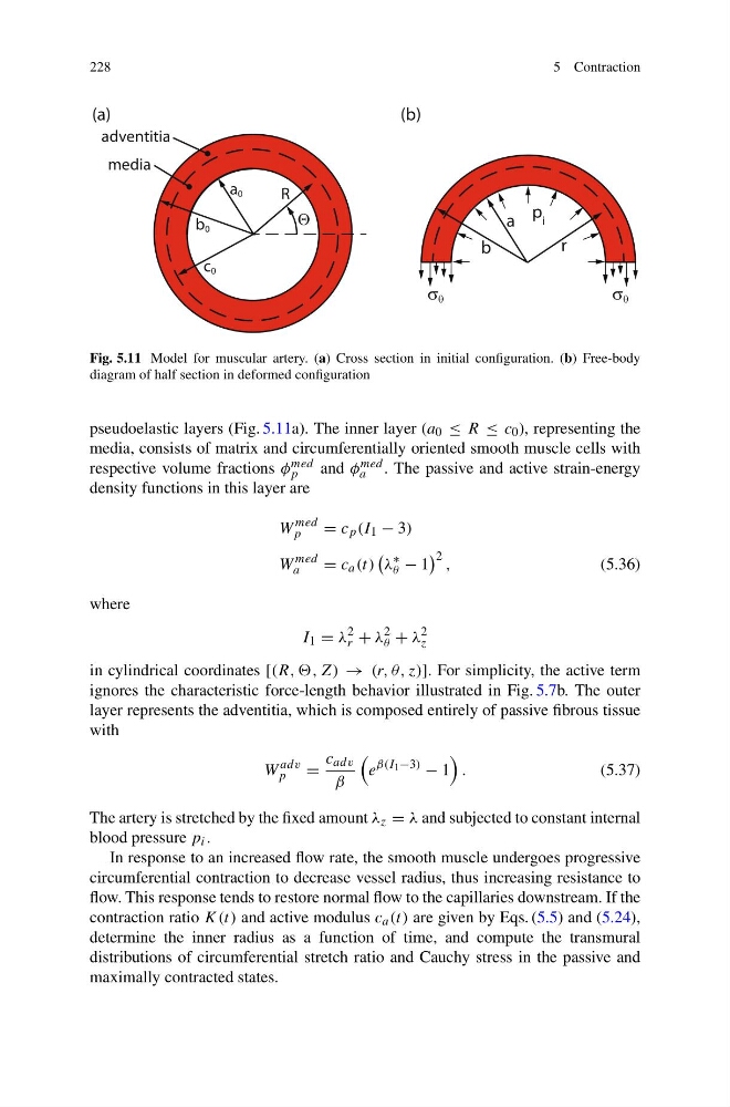
Fig. 5.11 Model for muscular artery. (a) Cross section in initial configuration. (b) Free-body
diagram of half section in deformed configuration
pseudoelastic layers (Fig. 5.11a). The inner layer (a 0 ≤ R ≤ c 0 ), representing the
media, consists of matrix and circumferentially oriented smooth muscle cells with
respective volume fractions φ med
p
and φ med
a . The passive and active strain-energy
density functions in this layer are
W
med
p
= c p (I 1 − 3)
W
med
a
= c a (t)
λ
∗
θ − 1
2 ,
(5.36)
where
I 1 = λ
2
r + λ
2
θ + λ
2
z
in cylindrical coordinates [(R, ,, Z) → (r, θ, z)]. For simplicity, the active term
ignores the characteristic force-length behavior illustrated in Fig. 5.7b. The outer
layer represents the adventitia, which is composed entirely of passive fibrous tissue
with
W
adv
p
=
c adv
β
e
β(I 1 −3)
− 1
.
(5.37)
The artery is stretched by the fixed amount λ z = λ and subjected to constant internal
blood pressure p i .
In response to an increased flow rate, the smooth muscle undergoes progressive
circumferential contraction to decrease vessel radius, thus increasing resistance to
flow. This response tends to restore normal flow to the capillaries downstream. If the
contraction ratio K(t) and active modulus c a (t) are given by Eqs. (5.5) and (5.24),
determine the inner radius as a function of time, and compute the transmural
distributions of circumferential stretch ratio and Cauchy stress in the passive and
maximally contracted states.
5 Contraction
R
Θ
a 0
c 0
b 0
adventitia
media
p i
)
b
(
)
a
(
σ θ
σ θ
a
r
b
Fig. 5.11 Model for muscular artery. (a) Cross section in initial configuration. (b) Free-body
diagram of half section in deformed configuration
pseudoelastic layers (Fig. 5.11a). The inner layer (a 0 ≤ R ≤ c 0 ), representing the
media, consists of matrix and circumferentially oriented smooth muscle cells with
respective volume fractions φ med
p
and φ med
a . The passive and active strain-energy
density functions in this layer are
W
med
p
= c p (I 1 − 3)
W
med
a
= c a (t)
λ
∗
θ − 1
2 ,
(5.36)
where
I 1 = λ
2
r + λ
2
θ + λ
2
z
in cylindrical coordinates [(R, ,, Z) → (r, θ, z)]. For simplicity, the active term
ignores the characteristic force-length behavior illustrated in Fig. 5.7b. The outer
layer represents the adventitia, which is composed entirely of passive fibrous tissue
with
W
adv
p
=
c adv
β
e
β(I 1 −3)
− 1
.
(5.37)
The artery is stretched by the fixed amount λ z = λ and subjected to constant internal
blood pressure p i .
In response to an increased flow rate, the smooth muscle undergoes progressive
circumferential contraction to decrease vessel radius, thus increasing resistance to
flow. This response tends to restore normal flow to the capillaries downstream. If the
contraction ratio K(t) and active modulus c a (t) are given by Eqs. (5.5) and (5.24),
determine the inner radius as a function of time, and compute the transmural
distributions of circumferential stretch ratio and Cauchy stress in the passive and
maximally contracted states.