

Ultrastructural studies of floral resin glands are scarce; it has been shown,
however, that the subcellular features of these glands are similar to those described
for resin glands in general [10, 13, 279, 284]. The predominant organelles are
plastids, mitochondria, and smooth endoplasmic reticulum, which are compatible
with the synthesis of terpene resins [10]. The epithelial cells of some species
of Clusia show all these characteristics and a prominent vacuole, sparse dictyosomes, and oil bodies inside plastids, small vacuoles, and vesicles free in the cytosol,
depending on the composition of the resin and its secretory pathway [279] (Fig. 18).
Plasmodesmata are not always present, and the production of vacuoles, in
some cases, is observed only during the secretory period, like in Philodendron
(Araceae) [13].
The mechanisms of resin release involve granulocrine and/or eccrine processes
(Fig. 18) [10, 11, 13, 56, 279]. Oil droplets and resin components can easily cross
the plasma membrane and cell walls, characterizing the eccrine process of secretion,
while some vesicles may fuse to the plasma membrane allowing some secretory
components to exit the cells through exocytosis, characterizing the secretory process
as granulocrine [279].
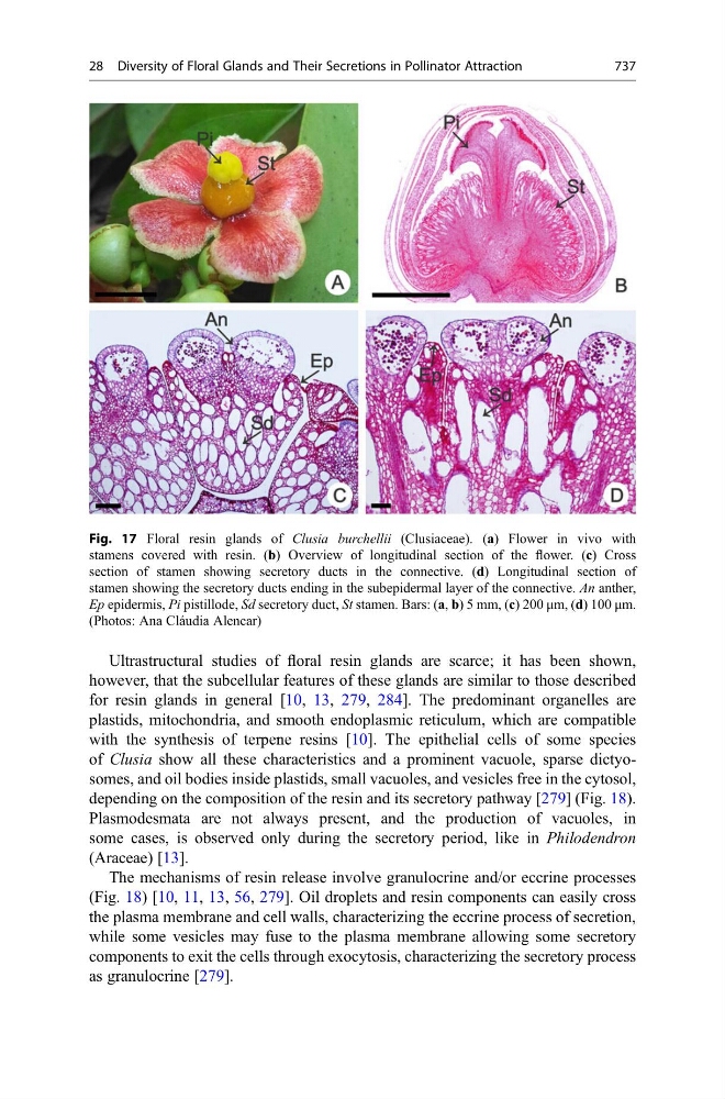
Fig. 17 Floral resin glands of Clusia burchellii (Clusiaceae). (a) Flower in vivo with
stamens covered with resin. (b) Overview of longitudinal section of the flower. (c) Cross
section of stamen showing secretory ducts in the connective. (d) Longitudinal section of
stamen showing the secretory ducts ending in the subepidermal layer of the connective. An anther,
Ep epidermis, Pi pistillode, Sd secretory duct, St stamen. Bars: (a, b) 5 mm, (c) 200 μm, (d) 100 μm.
(Photos: Ana Cláudia Alencar)
28 Diversity of Floral Glands and Their Secretions in Pollinator Attraction
737