

4.1
Structure and Location of Elaiophores
Anatomically, elaiophores are subdivided into two broad categories: epidermal and
trichomatous. Epidermal elaiophores consist of secretory epidermal cells, which are
generally elongate and accumulate the oil under their cuticles, forming “blisters” [11,
56, 216–218]. These secretory structures are documented in two families of eudicots,
Malpighiaceae and Krameriaceae, and two families of monocots, Orchidaceae and
only one species of Iridaceae [219–222]. Epidermal elaiophores are positioned on
tepals or petals (Fig. 13a) and/or sepals [223–228].
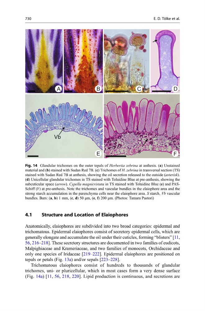
Trichomatous elaiophores consist of hundreds to thousands of glandular
trichomes, uni- or pluricellular, which in most cases form a very dense surface
(Fig. 14a) [11, 56, 218, 220]. Lipid production is continuous, and secretions are
Fig. 14 Glandular trichomes on the outer tepals of Herbertia zebrina at anthesis. (a) Unstained
material and (b) stained with Sudan Red 7B. (c) Trichomes of H. zebrina in transversal section (TS)
stained with Sudan Red 7B at anthesis, showing the oil secretion released to the outside (asterisk).
(d) Unicellular glandular trichomes in TS stained with Toluidine Blue at pre-anthesis, showing the
subcuticular space (arrow). Cypella magnicristata in TS stained with Toluidine Blue (e) and PASSchiff (F) at pre-anthesis. Note the trichomes and vascular bundles in the elaiophore area and the
strong starch accumulation in the parenchyma cells near the elaiophore area. S starch, Vb vascular
bundles. Bars: (a, b) 1 mm, (c, d) 50 μm, (e, f) 200 μm. (Photos: Tamara Pastori)
730
E. D. Tölke et al.