

separated from the reaction mixture using a magnetic field
and be used without any loss in the conversion rate.
Veisi et al. (2020) prepared a core–shell bionanocomposite to be used as a support for the production of Pd NPs.
For this aim, Fe 3 O 4 nanoparticles were prepared, and then
they were coated with aminopropyl silica. Finally, the prepared hybrid was functionalized with chitosan to introduce
different functional groups including hydroxyl and amine on
its surface. These functionalities provide a proper substrate
for coordination of metal ion and the prepared Pd nanoparticles will be anchored on the surface of the
chitosan-encapsulated Fe 3 O 4 /SiO 2 system. The fabricated
nanocomposite was applied in Suzuki–Miyaura coupling
reactions of a series of aryl halides and it showed superior
performance over other Pd-based catalysts. Also, its efficiency in catalyzing the reduction of 4-nitrophenol to
4-aminophenol was studied and satisfactory results were
obtained.
Abolghassem et al. (2019) used a-chitin as a template for
the synthesis of ZnO nanoparticles. In this procedure, ZnO
nanoparticles were produced in situ during a hydrothermal
process and then the prepared chitin/ZnO nanocomposite
was used as a catalyst for the synthesis of benzo[a]pyrano(2,
3-c)phenazine derivatives. This green manner was proposed
as an efficient way to produce these pharmaceutically
important materials in satisfactory yields.
A blend composed of chitosan and PVA was used with
dual function to provide both carbon and nitrogen in the
preparation of Ag nanoparticles embedded with
nitrogen-doped carbon nanocomposite (Alhokbany et al.
2019). Figure 17 shows the related procedure, schematically. The prepared nitrogen-doped graphite carbon matrix
affords a bed for not only proper dispersion of Ag
nanoparticles, but also enhances its recyclability and consequently its life time. The catalytic efficiency of the prepared nanocomposites with relatively supreme surface area
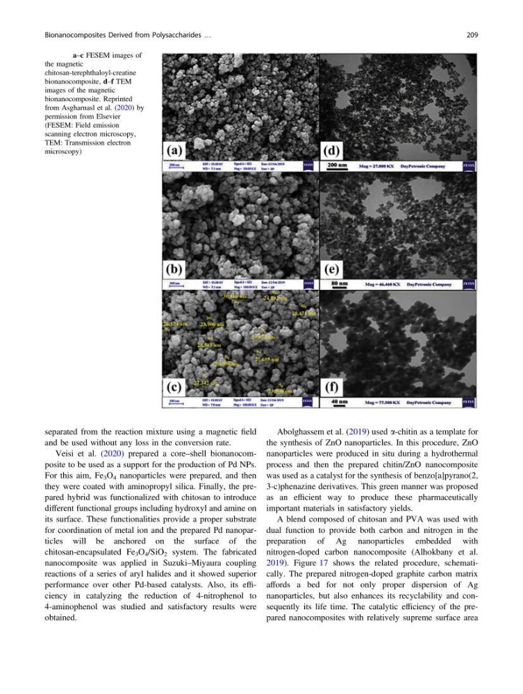
Fig. 15 a–c FESEM images of
the magnetic
chitosan-terephthaloyl-creatine
bionanocomposite, d–f TEM
images of the magnetic
bionanocomposite. Reprinted
from Asgharnasl et al. (2020) by
permission from Elsevier
(FESEM: Field emission
scanning electron microscopy,
TEM: Transmission electron
microscopy)
Bionanocomposites Derived from Polysaccharides …
209
and be used without any loss in the conversion rate.
Veisi et al. (2020) prepared a core–shell bionanocomposite to be used as a support for the production of Pd NPs.
For this aim, Fe 3 O 4 nanoparticles were prepared, and then
they were coated with aminopropyl silica. Finally, the prepared hybrid was functionalized with chitosan to introduce
different functional groups including hydroxyl and amine on
its surface. These functionalities provide a proper substrate
for coordination of metal ion and the prepared Pd nanoparticles will be anchored on the surface of the
chitosan-encapsulated Fe 3 O 4 /SiO 2 system. The fabricated
nanocomposite was applied in Suzuki–Miyaura coupling
reactions of a series of aryl halides and it showed superior
performance over other Pd-based catalysts. Also, its efficiency in catalyzing the reduction of 4-nitrophenol to
4-aminophenol was studied and satisfactory results were
obtained.
Abolghassem et al. (2019) used a-chitin as a template for
the synthesis of ZnO nanoparticles. In this procedure, ZnO
nanoparticles were produced in situ during a hydrothermal
process and then the prepared chitin/ZnO nanocomposite
was used as a catalyst for the synthesis of benzo[a]pyrano(2,
3-c)phenazine derivatives. This green manner was proposed
as an efficient way to produce these pharmaceutically
important materials in satisfactory yields.
A blend composed of chitosan and PVA was used with
dual function to provide both carbon and nitrogen in the
preparation of Ag nanoparticles embedded with
nitrogen-doped carbon nanocomposite (Alhokbany et al.
2019). Figure 17 shows the related procedure, schematically. The prepared nitrogen-doped graphite carbon matrix
affords a bed for not only proper dispersion of Ag
nanoparticles, but also enhances its recyclability and consequently its life time. The catalytic efficiency of the prepared nanocomposites with relatively supreme surface area
Fig. 15 a–c FESEM images of
the magnetic
chitosan-terephthaloyl-creatine
bionanocomposite, d–f TEM
images of the magnetic
bionanocomposite. Reprinted
from Asgharnasl et al. (2020) by
permission from Elsevier
(FESEM: Field emission
scanning electron microscopy,
TEM: Transmission electron
microscopy)
Bionanocomposites Derived from Polysaccharides …
209