

4.9 Outcome of Characterization
85
in the crystallinity of GXCS. This observation is due to the fact that some of the
crystalline chains have been eliminated in the process of grafting.
4.9.2 Outcome of SEM Morphology Investigation
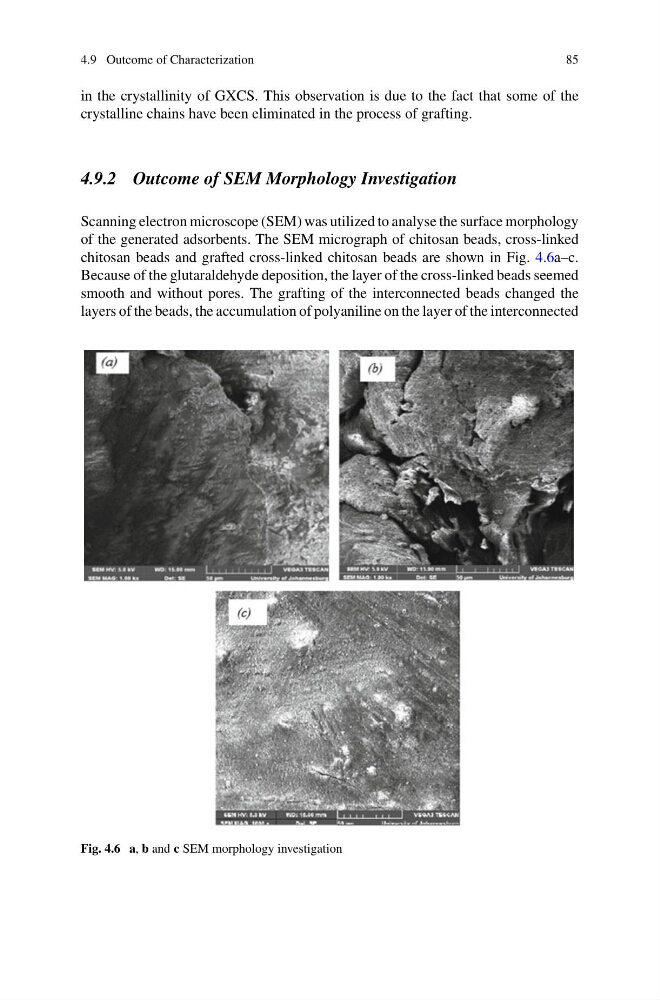
Scanning electron microscope (SEM) was utilized to analyse the surface morphology
of the generated adsorbents. The SEM micrograph of chitosan beads, cross-linked
chitosan beads and grafted cross-linked chitosan beads are shown in Fig. 4.6a–c.
Because of the glutaraldehyde deposition, the layer of the cross-linked beads seemed
smooth and without pores. The grafting of the interconnected beads changed the
layers of the beads, the accumulation of polyaniline on the layer of the interconnected
Fig. 4.6 a, b and c SEM morphology investigation
85
in the crystallinity of GXCS. This observation is due to the fact that some of the
crystalline chains have been eliminated in the process of grafting.
4.9.2 Outcome of SEM Morphology Investigation
Scanning electron microscope (SEM) was utilized to analyse the surface morphology
of the generated adsorbents. The SEM micrograph of chitosan beads, cross-linked
chitosan beads and grafted cross-linked chitosan beads are shown in Fig. 4.6a–c.
Because of the glutaraldehyde deposition, the layer of the cross-linked beads seemed
smooth and without pores. The grafting of the interconnected beads changed the
layers of the beads, the accumulation of polyaniline on the layer of the interconnected
Fig. 4.6 a, b and c SEM morphology investigation