

number of scientific communities. In particular, for life science communities, it is
available as the Fiji plug-in platform, which allows for easy plug-in deployment and
dependency management. ImageJ has an open architecture providing extensibility
via third-party Java modules (called plug-ins) and scripting macros. It is developed
by Wayne Rasband since 1997 and expanded via contributed software code by an
international group of contributors. Plug-ins are distributed together with their
source code under various licenses determined by the plug-in authors. The user
guide of the platform [23] is maintained at http://imagej.nih.gov/ij/docs/guide.
Public resources are available on the ImageJ website and the ImageJ Information
and Documentation Portal https://imagejdocu.list.lu/. In addition, textbook
introductions to image processing with ImageJ can be found in [24].
7. Discussion
The morphological complexity of the nervous tissue is a challenge for conventional segmentation techniques developed for computer vision applications or cultured cells. The challenges lie in the morphological complexity of neurons and glial
cells overlaid on the heterogeneity of the extracellular matrix. This complexity
translates into variations of the tracer signal and touching of relevant structures.
Segmentation of fluorescent images poses particular issues due to low signal-tonoise ratio, unequal staining, as well as the complexity of structures that need to be
identified. This irreducible variation must inform choices about segmentation
methods. In particular, methods employing multiple spatial scales are favorable.
Structure identification is inherently a multiscale problem because object structure
is recursive, that is, objects may contain substructures, which themselves contain
substructures, etc.
A large number of algorithms for image segmentation have been proposed in
literature (overview in [9]). However, many of them completely ignore the issue of
scale. As a result, they are capable of identifying only limited types of structures. In
contrast, multiscale approaches eventually rely on the topological properties of the
segmented objects, either by means of scale spaces or by nonlinear vector field
transforms [25, 26]. As a result, such methods are able to combine detected features
into robust segmentation tools. The present chapter introduced two classes of
multiscale methods for image segmentation: the mathematical morphology operations and scale spaces. The main applications of the theory are classification and
segmentation of signals. Presented methods are generic and thus have broad applicability to both one-dimensional signals, such as electrophysiological recordings,
and to two and three-dimensional signals, such as microscopic images.
Plug-in
Function
LoG filter
Laplacian of Gaussian (LoG)
ALoG filter
Anisotropic decomposition of LoG
ADiff filter
Anisotropic diffusion
LoG2 filter
Bi-Laplacian of Gaussian
LoGN2D filter
N-order power of the Laplacian of Gaussian
Gaussian jet
Gaussian jet of order n
Zero-crosser
Connected components
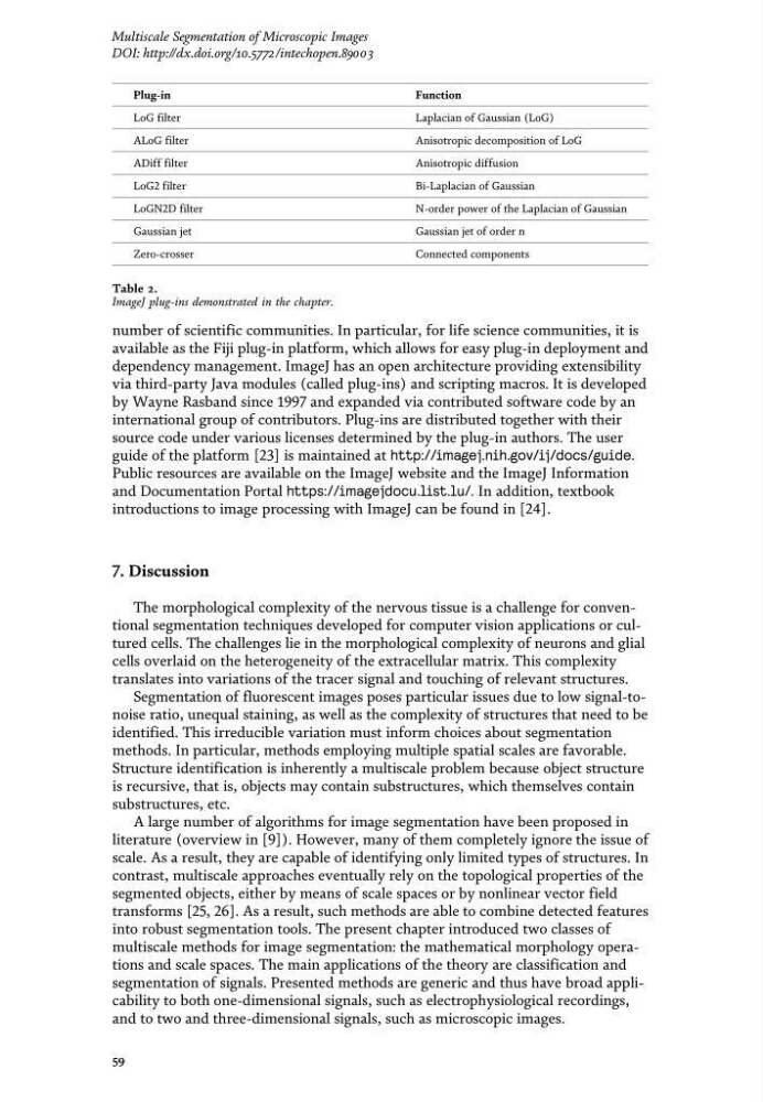
Table 2.
ImageJ plug-ins demonstrated in the chapter.
59
Multiscale Segmentation of Microscopic Images
DOI: http://dx.doi.org/10.5772/intechopen.89003
available as the Fiji plug-in platform, which allows for easy plug-in deployment and
dependency management. ImageJ has an open architecture providing extensibility
via third-party Java modules (called plug-ins) and scripting macros. It is developed
by Wayne Rasband since 1997 and expanded via contributed software code by an
international group of contributors. Plug-ins are distributed together with their
source code under various licenses determined by the plug-in authors. The user
guide of the platform [23] is maintained at http://imagej.nih.gov/ij/docs/guide.
Public resources are available on the ImageJ website and the ImageJ Information
and Documentation Portal https://imagejdocu.list.lu/. In addition, textbook
introductions to image processing with ImageJ can be found in [24].
7. Discussion
The morphological complexity of the nervous tissue is a challenge for conventional segmentation techniques developed for computer vision applications or cultured cells. The challenges lie in the morphological complexity of neurons and glial
cells overlaid on the heterogeneity of the extracellular matrix. This complexity
translates into variations of the tracer signal and touching of relevant structures.
Segmentation of fluorescent images poses particular issues due to low signal-tonoise ratio, unequal staining, as well as the complexity of structures that need to be
identified. This irreducible variation must inform choices about segmentation
methods. In particular, methods employing multiple spatial scales are favorable.
Structure identification is inherently a multiscale problem because object structure
is recursive, that is, objects may contain substructures, which themselves contain
substructures, etc.
A large number of algorithms for image segmentation have been proposed in
literature (overview in [9]). However, many of them completely ignore the issue of
scale. As a result, they are capable of identifying only limited types of structures. In
contrast, multiscale approaches eventually rely on the topological properties of the
segmented objects, either by means of scale spaces or by nonlinear vector field
transforms [25, 26]. As a result, such methods are able to combine detected features
into robust segmentation tools. The present chapter introduced two classes of
multiscale methods for image segmentation: the mathematical morphology operations and scale spaces. The main applications of the theory are classification and
segmentation of signals. Presented methods are generic and thus have broad applicability to both one-dimensional signals, such as electrophysiological recordings,
and to two and three-dimensional signals, such as microscopic images.
Plug-in
Function
LoG filter
Laplacian of Gaussian (LoG)
ALoG filter
Anisotropic decomposition of LoG
ADiff filter
Anisotropic diffusion
LoG2 filter
Bi-Laplacian of Gaussian
LoGN2D filter
N-order power of the Laplacian of Gaussian
Gaussian jet
Gaussian jet of order n
Zero-crosser
Connected components
Table 2.
ImageJ plug-ins demonstrated in the chapter.
59
Multiscale Segmentation of Microscopic Images
DOI: http://dx.doi.org/10.5772/intechopen.89003